CXR findings, key signs and common patterns
Pulmonary oedema is something every medical professional needs to recognise. Chest X-rays are quick and widely available, but the findings can be subtle and easy to miss if you’re not sure what to look for.
In this article, we walk through a real case and break down the key imaging features step by step. You’ll learn how to spot classic signs like Kerley B lines and the bat wing pattern, understand why fluid builds up in the lungs, and use the ABCDE approach to interpret X-rays with more confidence.
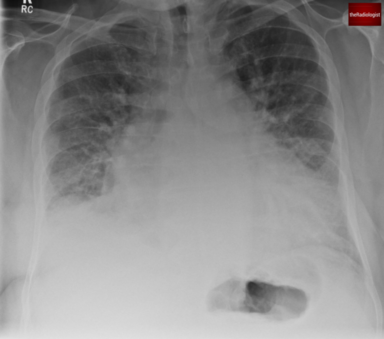
A 70-year-old male presents with breathlessness: have a look at his PA chest X-Ray. Let’s go through the case and have a think about pulmonary oedema.
PA view of a chest X-Ray of a male in his 70s
Here is a video explanation of this case: click full screen in the bottom right corner to make it big. If you prefer though I go through this in the text explanation below.
Pulmonary oedema is the accumulation of extravascular fluid in the lungs. This can occur due to various mechanisms, but the most common cause is cardiogenic pulmonary oedema, often stemming from left heart failure. Non-cardiogenic causes include:
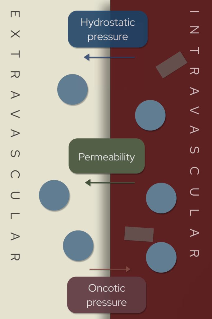
How does this extravascular fluid build up?
Three key factors govern the movement of fluid between capillaries and interstitial spaces:
Increased hydrostatic pressure, increased permeability and reduced oncotic pressure can all lead to increased extravascular fluid and pulmonary oedema.
Pulmonary oedema has a classic appearance on chest X-Ray. Let’s explore the key findings:
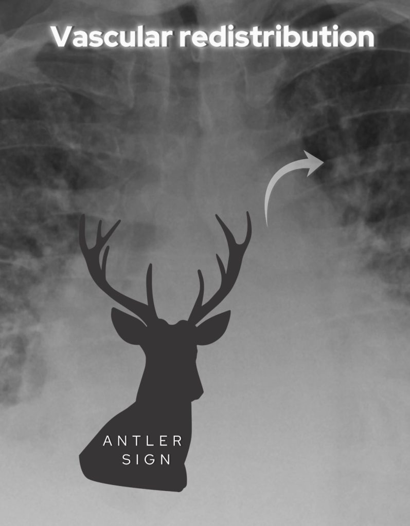
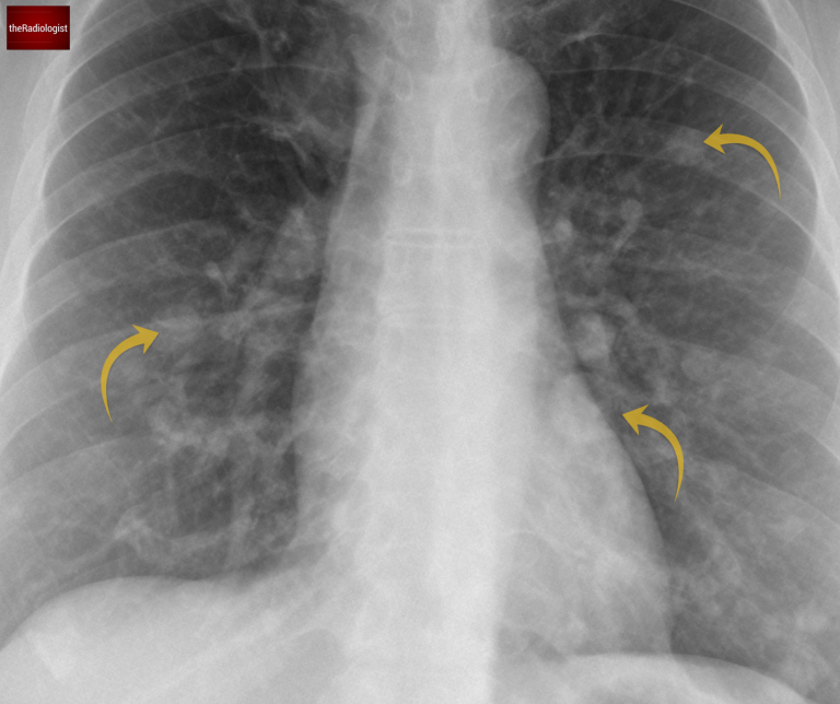
Look for dilated upper lobe veins – the ‘antler’ sign
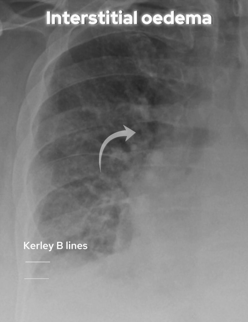
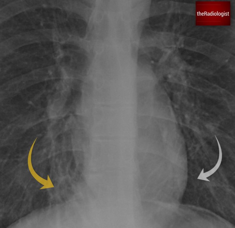
Look for thickening of bronchial walls and loss of definition of the airways. Kerley B lines were not present in this case but are seen as horizontal lines at the lung base reflecting interstitial thickening.
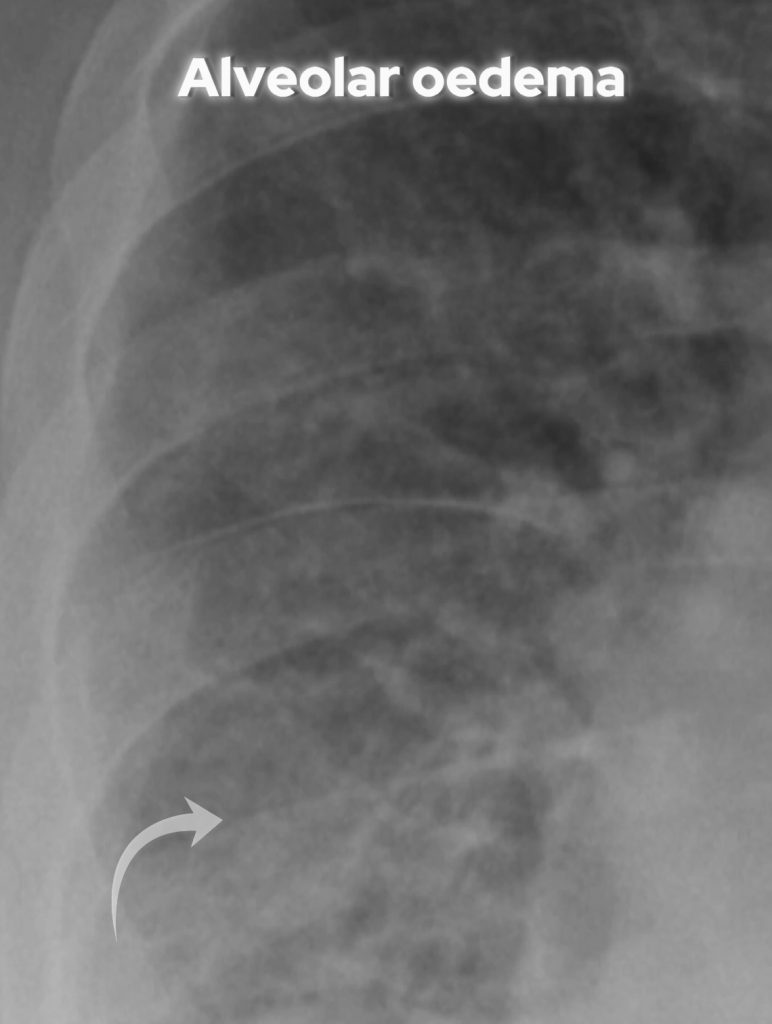
Look for ‘fluffy’ alveolar oedema
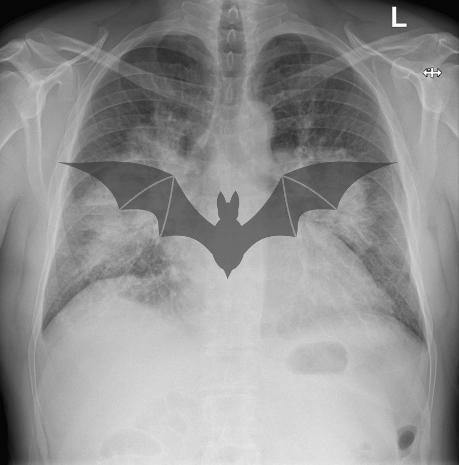
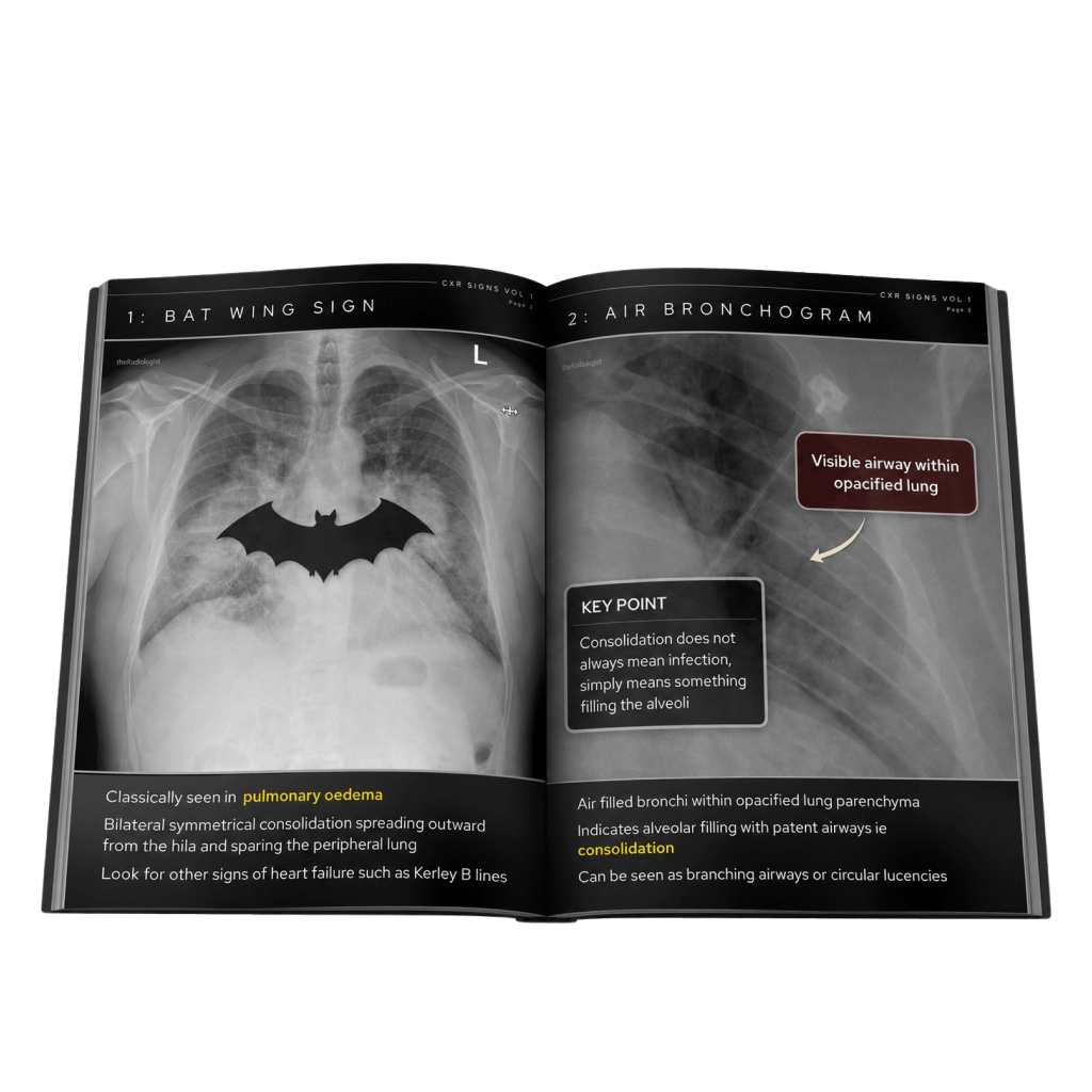
In this separate case there is rapid fluid accumulation leading to a classic bat wing appearance of pulmonary oedema.
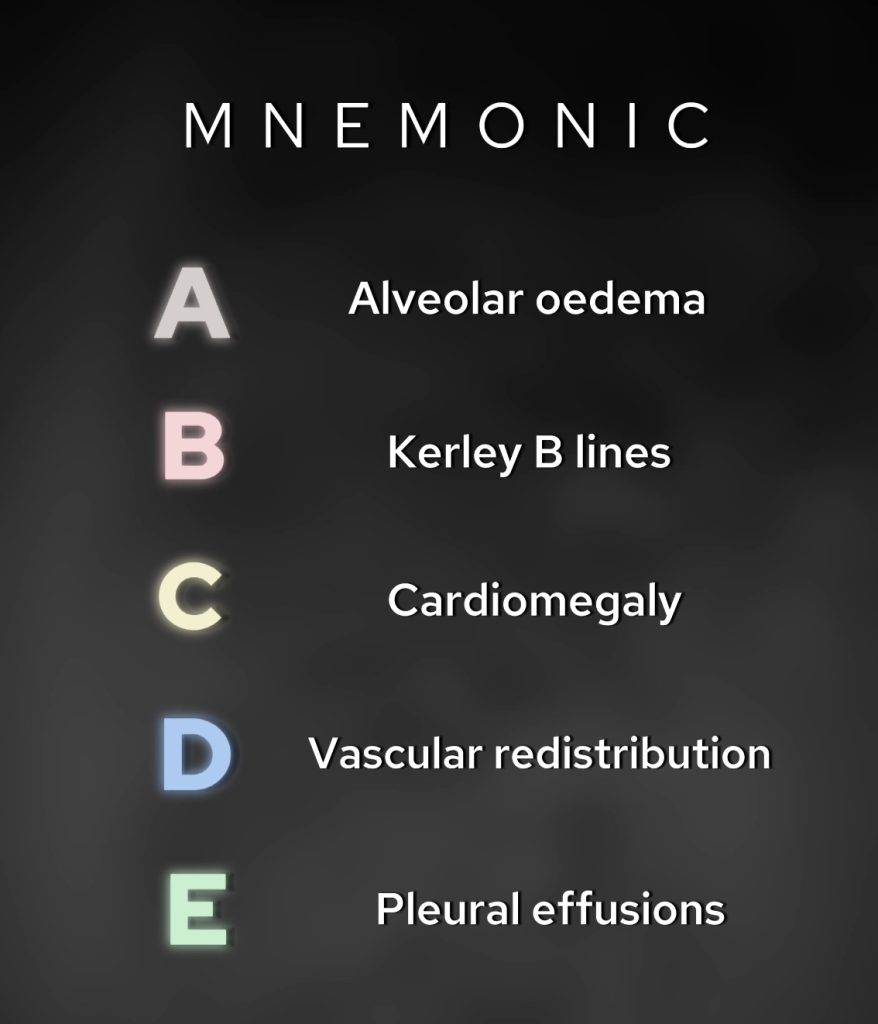
Here’s a handy mnemonic I’ve remembered since my days at med school about the findings of pulmonary oedema on chest X-Ray:
In this patient, the findings are consistent with cardiogenic pulmonary oedema, caused by left ventricular dysfunction secondary to ischaemic heart disease.
We see vascular redistribution, interstitial oedema, and alveolar oedema on the X-ray.
Pulmonary oedema can be classified into four categories based on the underlying mechanism:
Don’t forget the ABCDE mnemonic for signs of pulmonary oedema – this is something I have been using since medical school and still use it to this day as a Consultant Radiologist.
Remember pulmonary oedema can be treated quickly and X-Ray features can resolve quickly (unless pneumonia and ARDS) so a short interval follow up X-Ray can help differentiate if things are difficult to interpret clinically.
Downloaded by 10,000+ healthcare professionals.
Get 20 annotated chest X-ray signs with clear teaching points and explanations. Written by a Consultant Radiologist, this free guide is designed to help you recognise important findings more confidently when reviewing chest X-Rays.
You’ve got the framework now let’s put it to work. Dive into a related case to see these findings on real images, or explore another guide to build out your systematic approach. That’s where it really starts to stick.
A systematic approach to anatomy, lines and tubes and review areas
Learn the X-Ray and CT features and aetiology of bronchiectasis
Chest X-ray and CT appearances, causes and differential diagnosis
We use cookies to make this website work and to understand how it is used. You can accept all cookies, use essential cookies only or manage your preferences.