
Anatomy gallery
Explore a wide range of annotated radiology images
Introduction
The Radiologist Anatomy Gallery is a collection of annotated radiology anatomy images, including labelled X-rays, CT, MRI and ultrasound examples. These guides are designed to help doctors, medical students, radiographers and healthcare professionals learn practical radiographic anatomy. These images are provided for personal learning only and must not be reused or shared without permission.
Upper limb
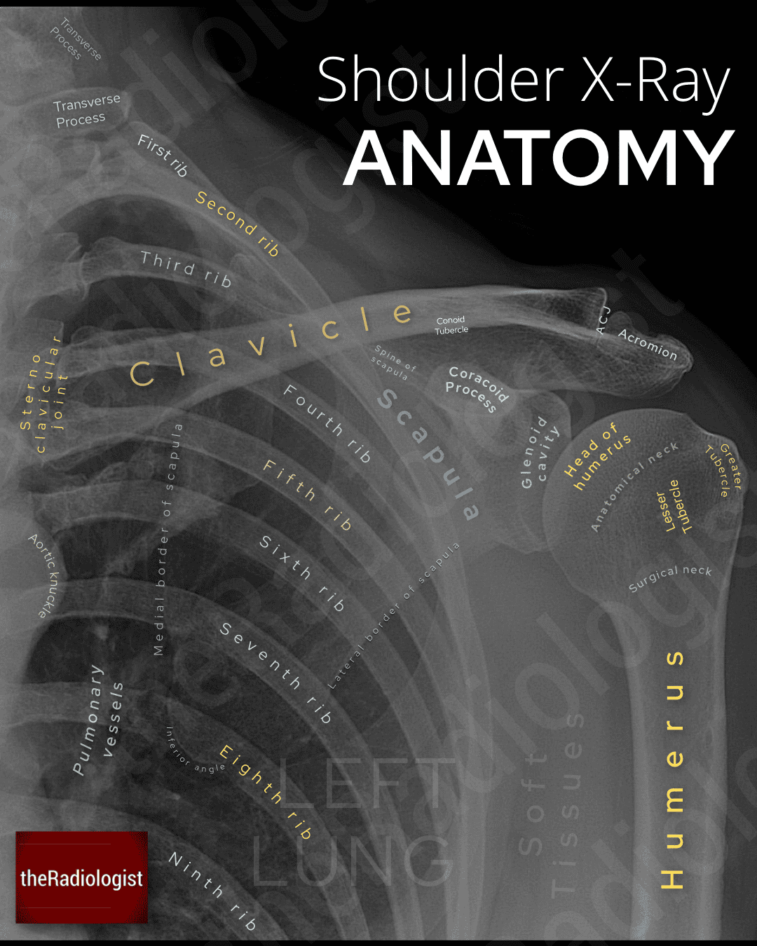
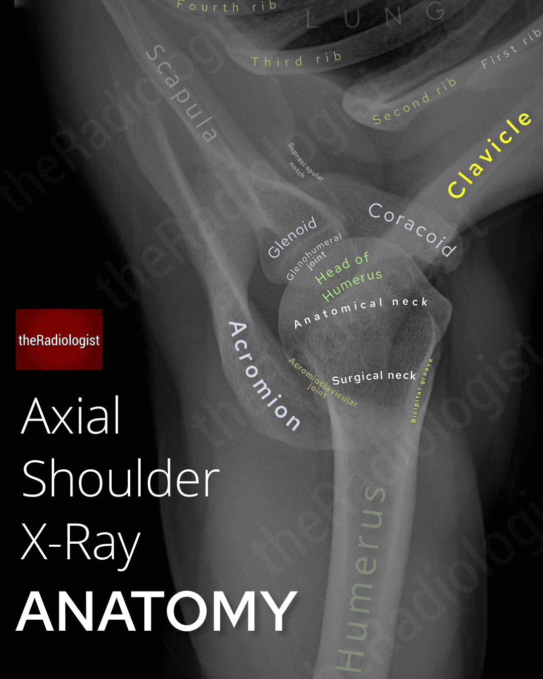
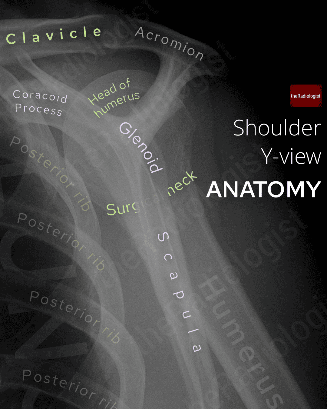
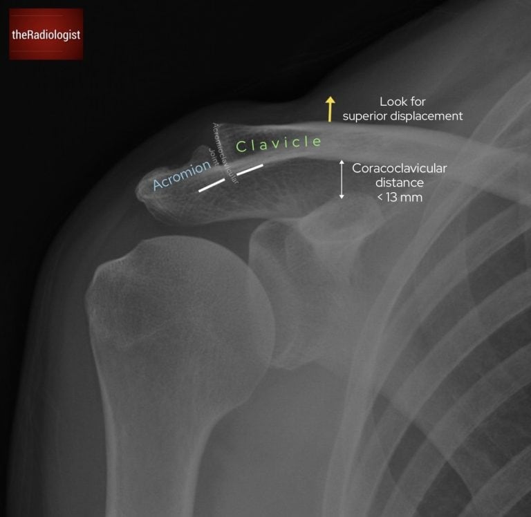
Shoulder X-Ray
The AP view forms the ‘primary view’ with other views such as the Y view or axial view being adjuncts – particularly in the acute setting where these adjuncts can help assess for glenohumeral dislocation.
→ Learn a review system to help go through shoulder X-Rays here.
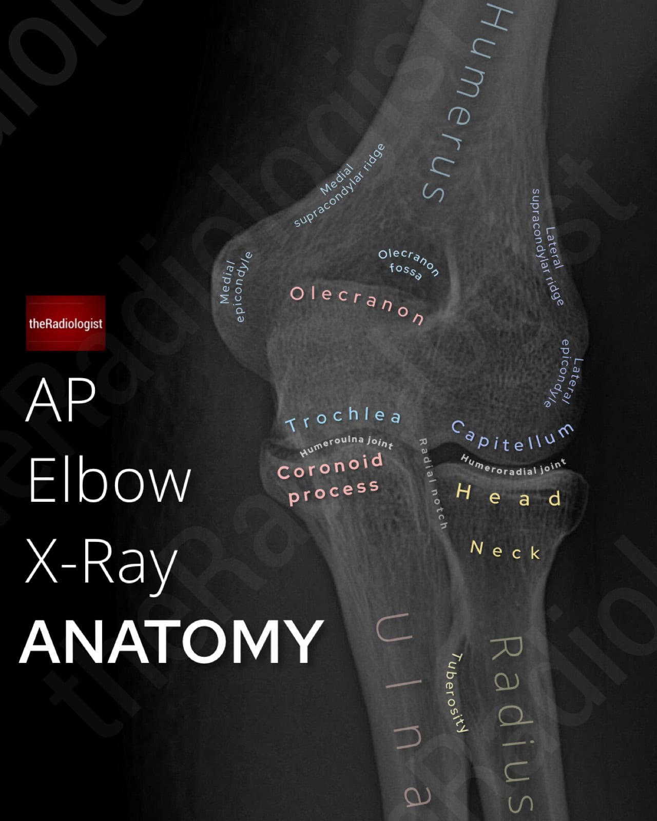
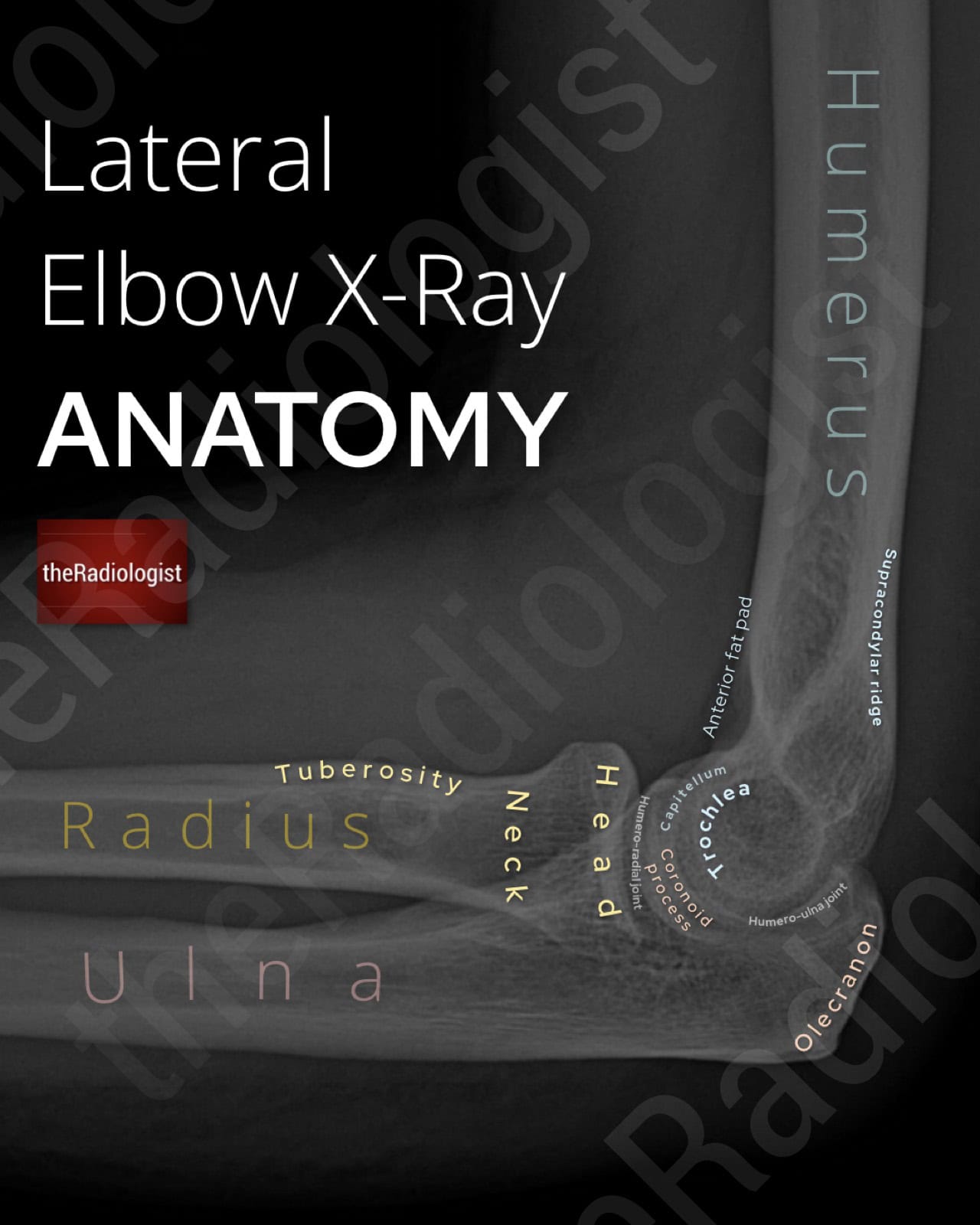
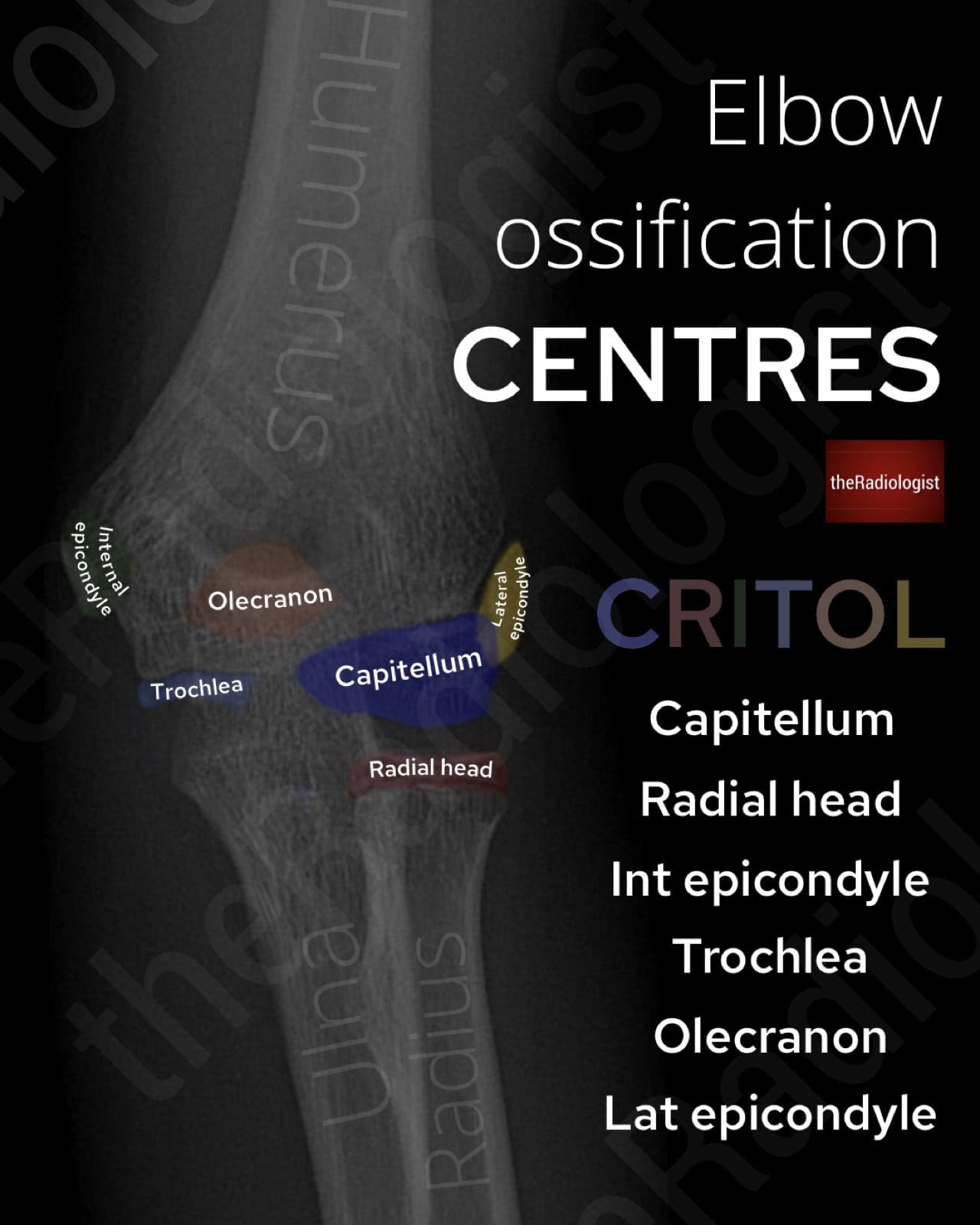
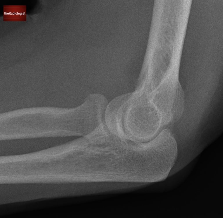
Elbow X-Ray
The main elbow views are the AP and lateral.
→ Go through the detail and learn a review system to assess these X-Rays here.
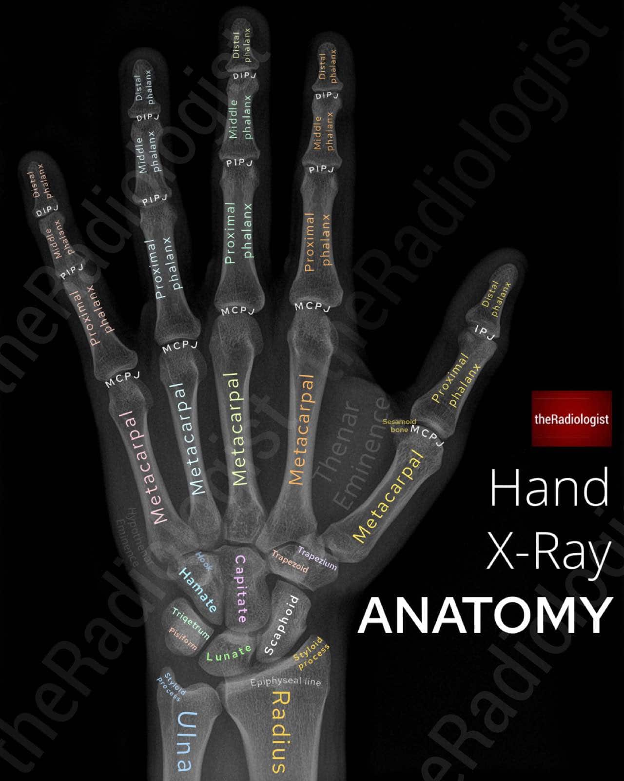
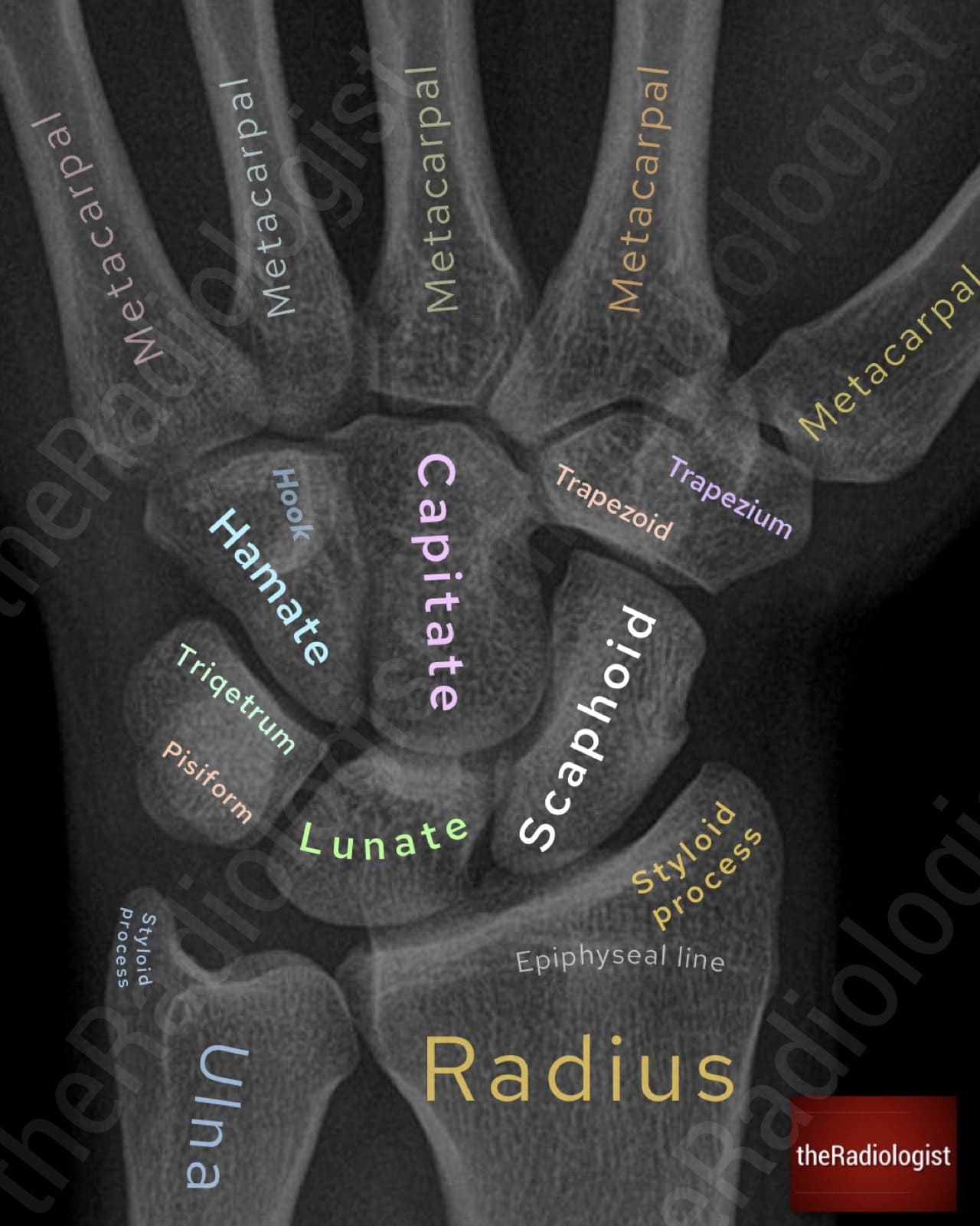
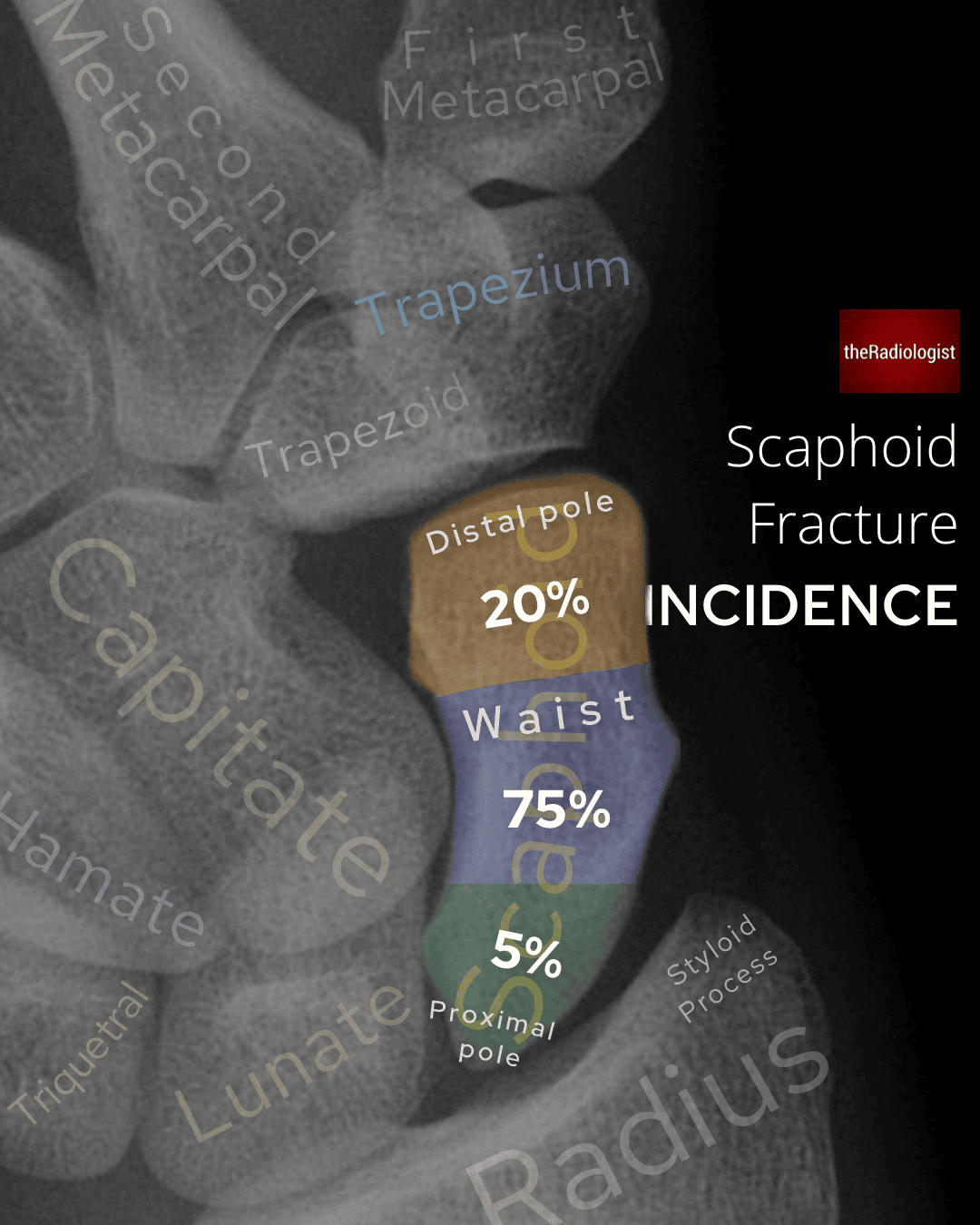
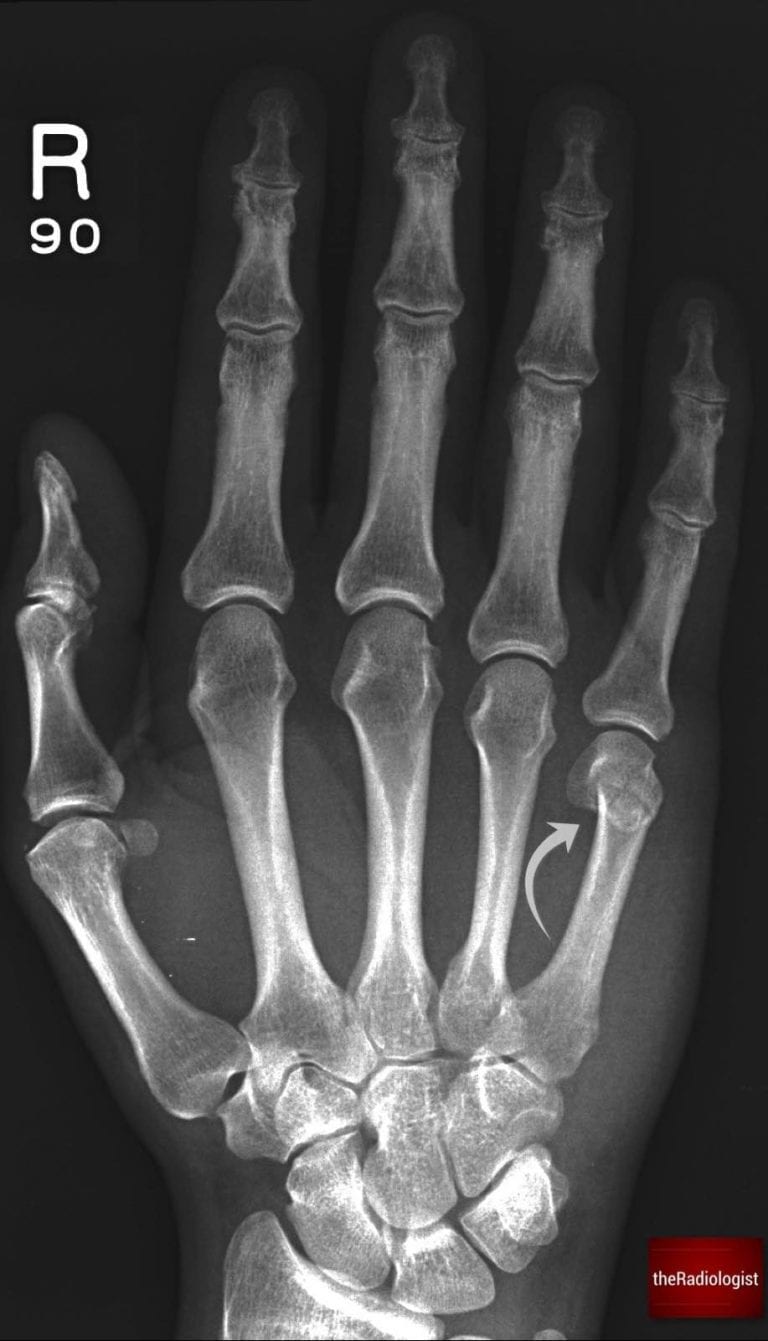
Hand and wrist X-Ray
Here we have a PA hand X-Ray as well as a PA view of the carpal bones.
→ Go through the detail and learn a review system to assess these X-Rays here.
Lower limb
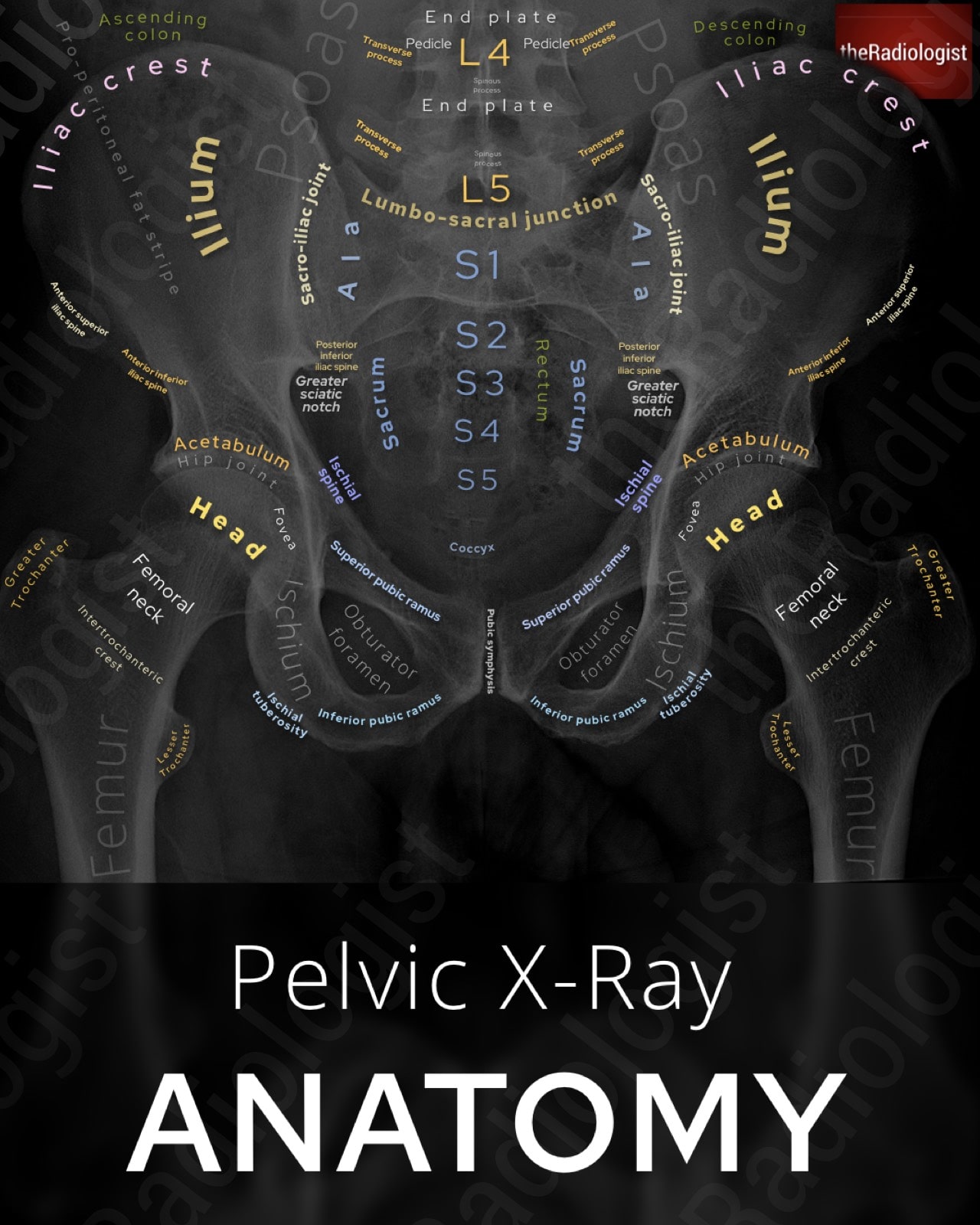
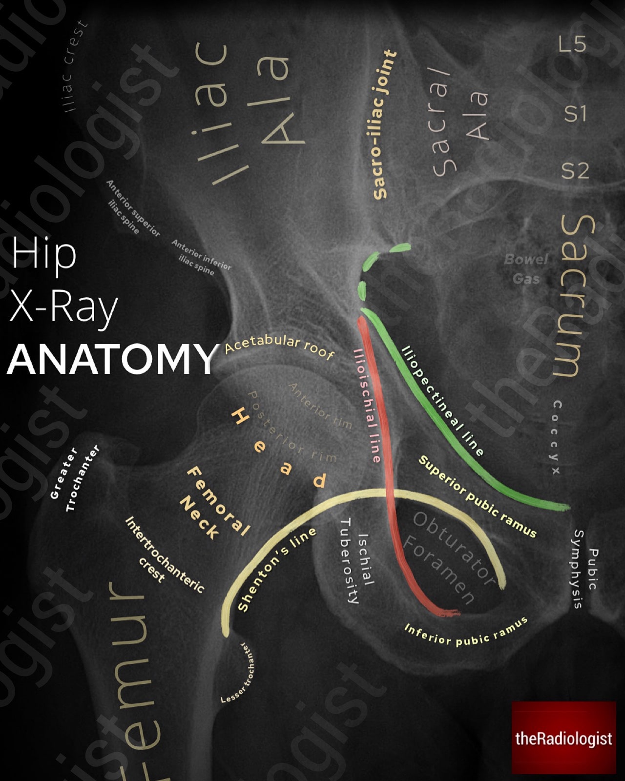
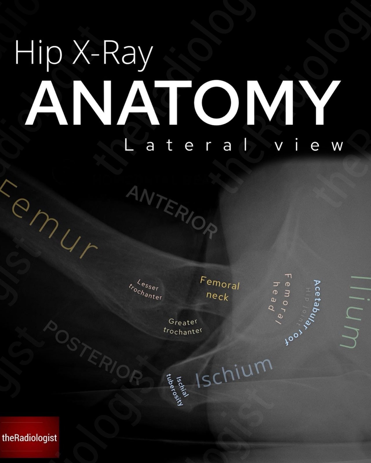
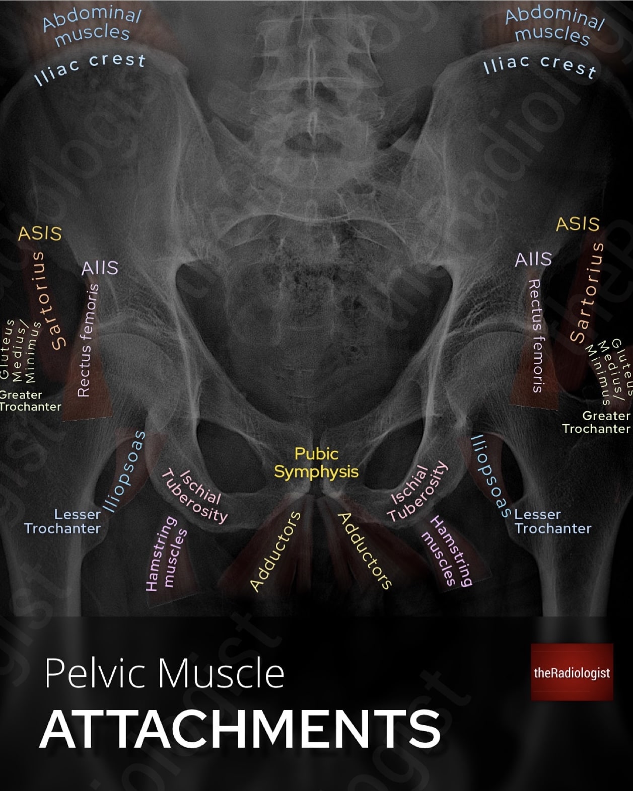
Pelvic X-Ray
Below we have an AP pelvic X-Ray, AP hip X-Ray and lateral hip X-Ray as well as a diagram of pelvic muscle attachments.
→ Go through the detail and learn a review system to assess these X-Rays here.
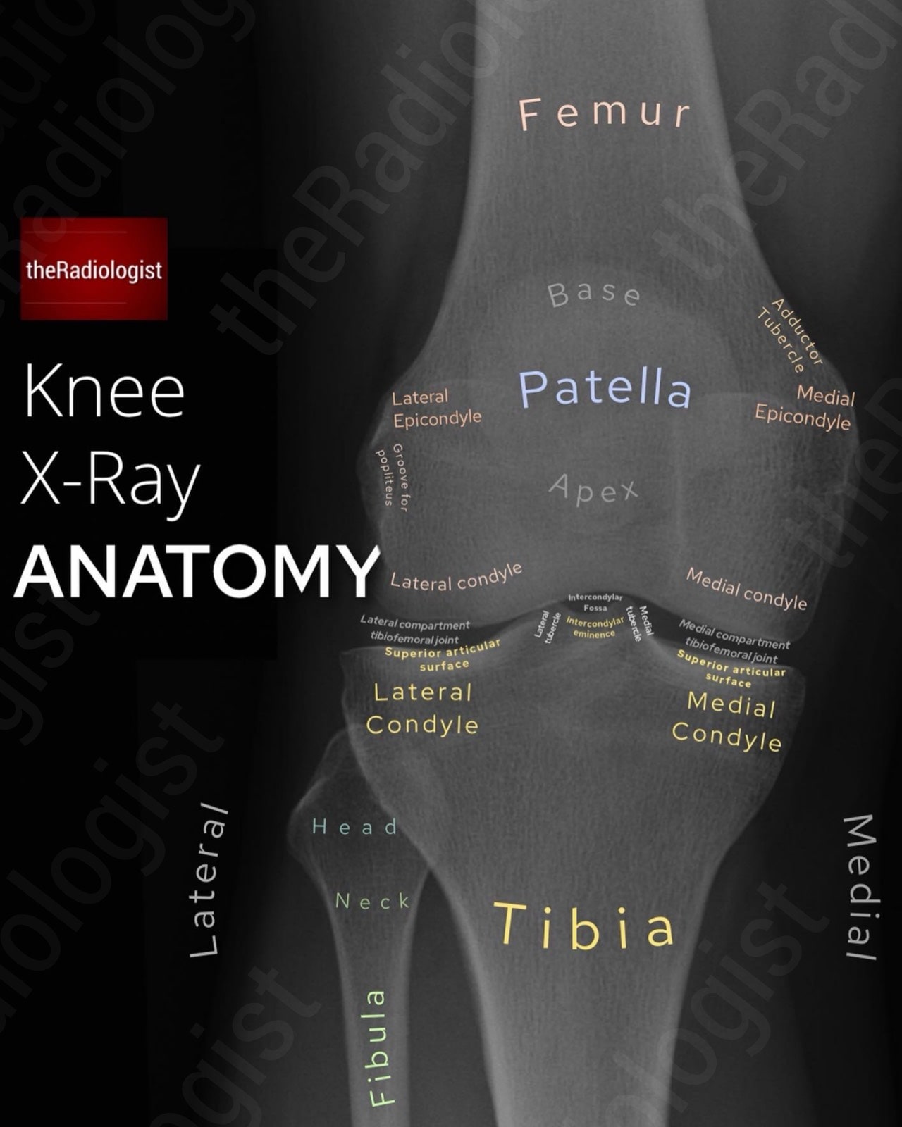
Knee X-Ray
Below we have annotated AP and lateral (HBL) views of a knee X-Ray.
→ Go through the detail and learn a review system to assess these X-Rays here.
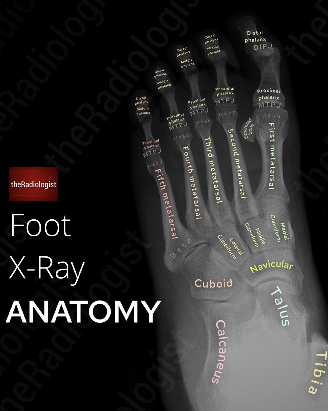
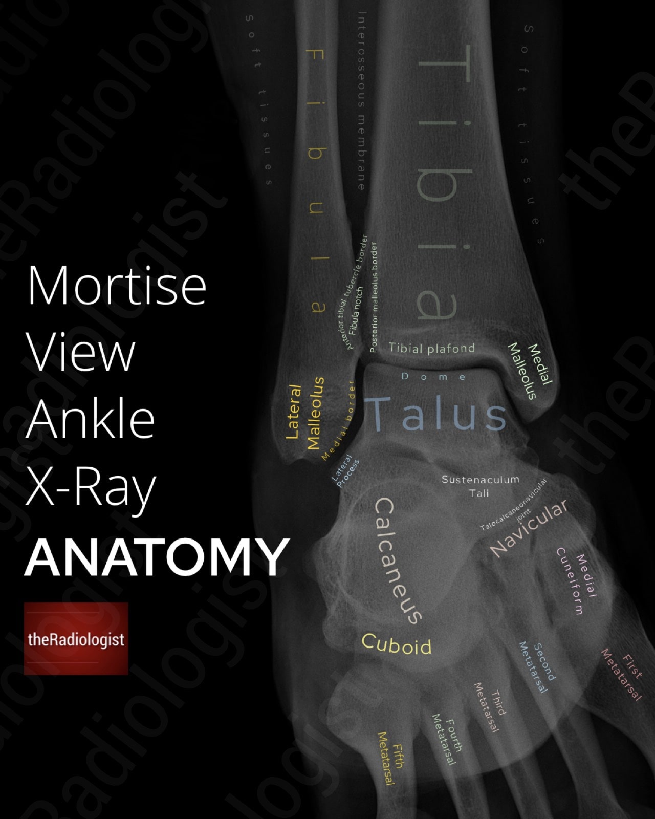
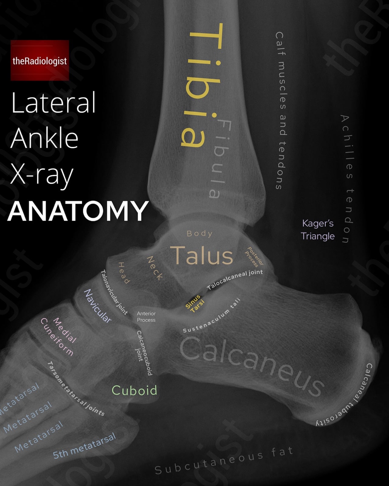
Foot and ankle X-Ray
Below we have annotated views of an oblique foot, mortise ankle and lateral ankle X-Ray.
→ Go through the detail and learn a review system to assess these X-Rays here.
Body
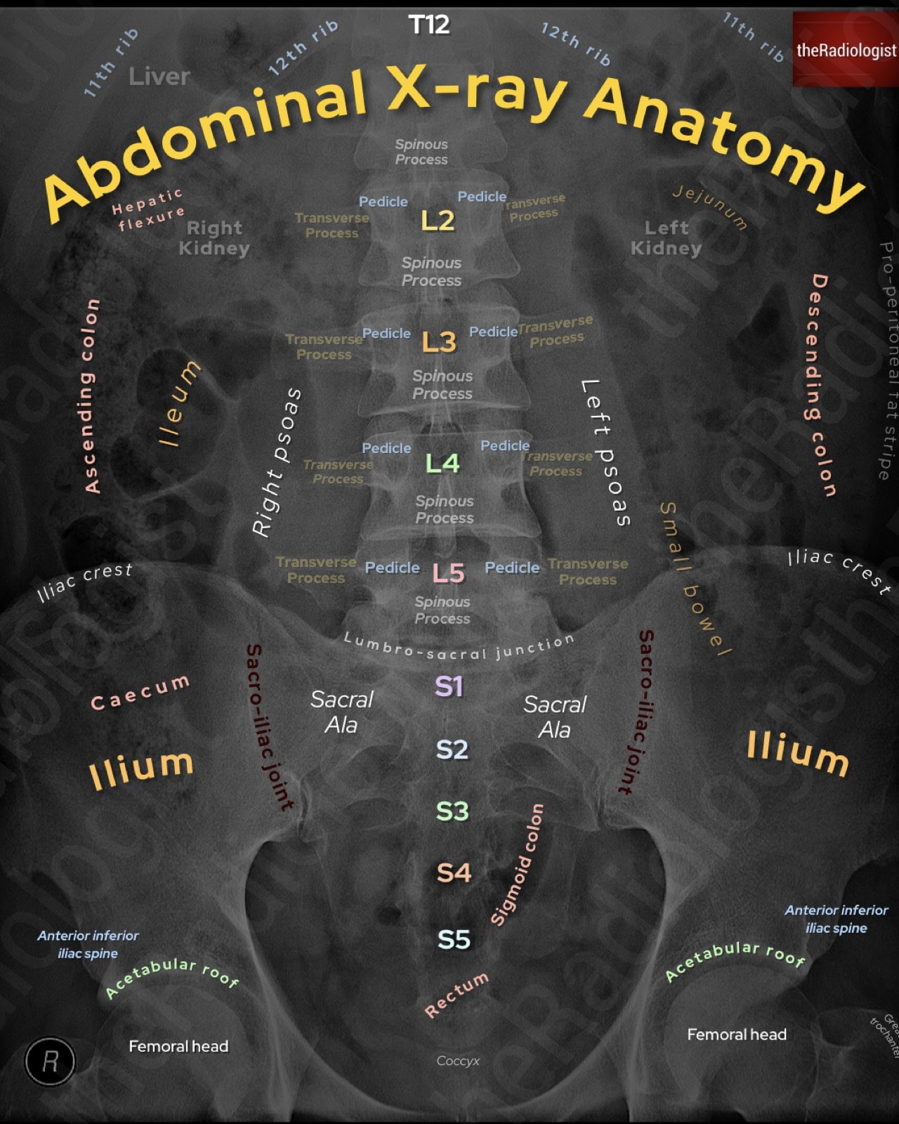
Abdominal X-Ray
Below we have an annotated view of an abdominal X-Ray.
→ Go through the detail and learn a review system to assess these X-Rays here.
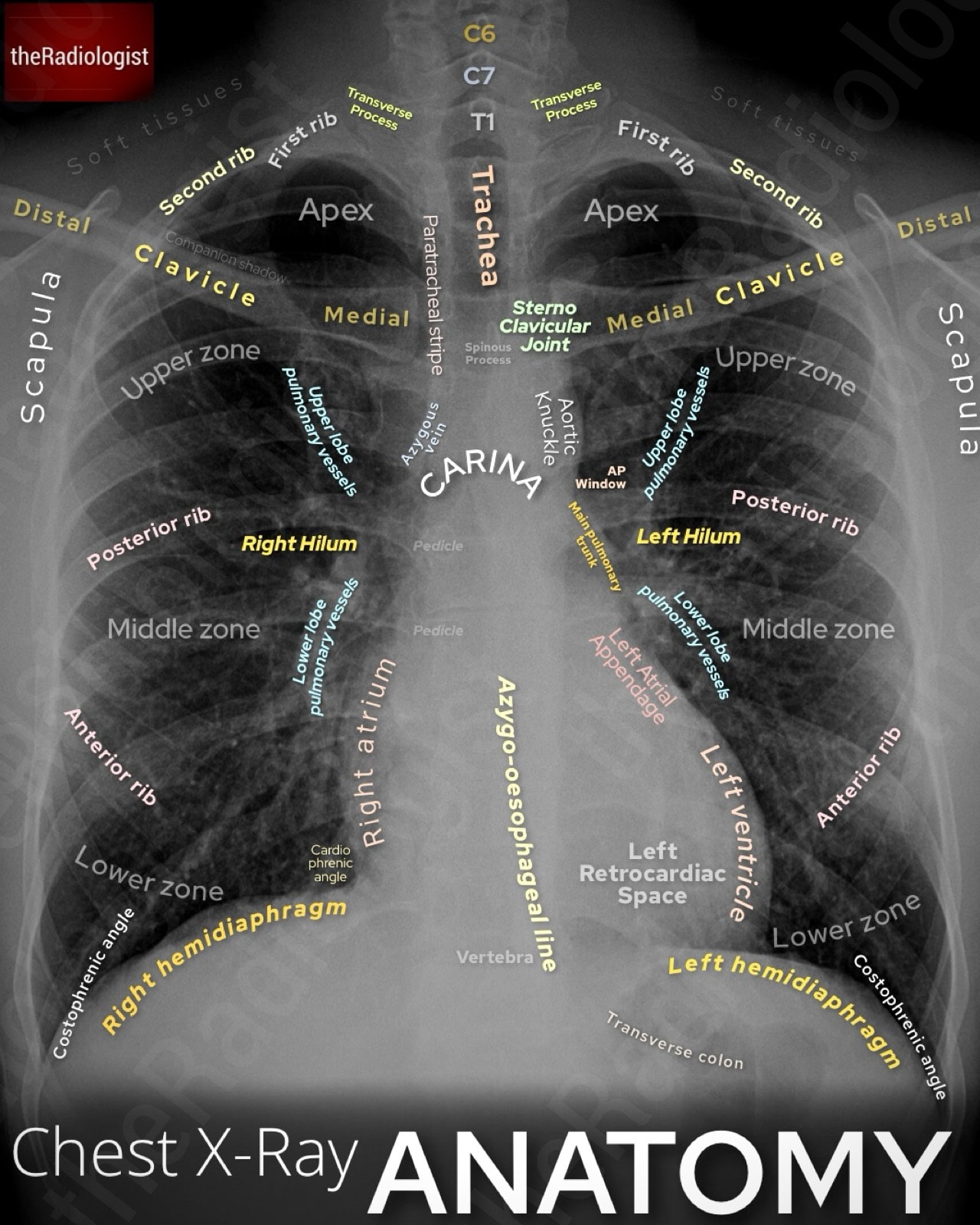
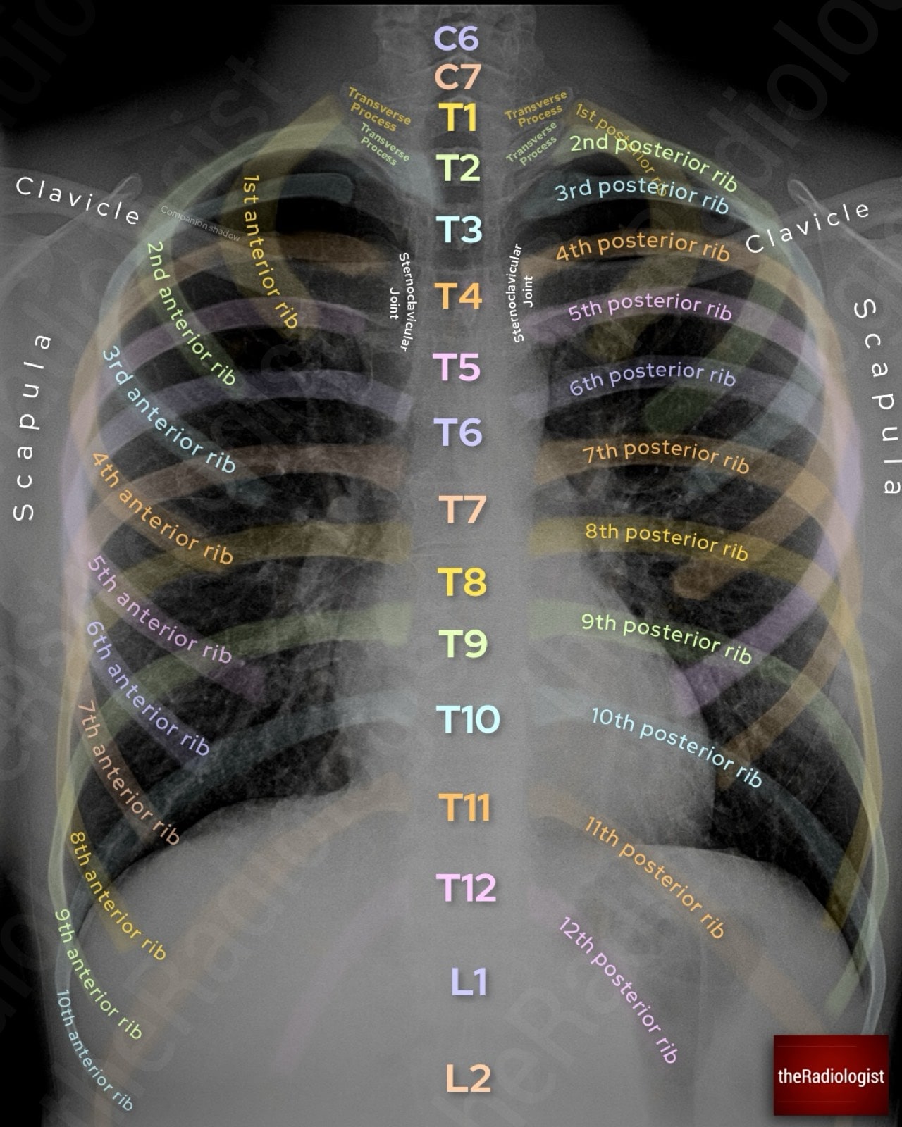
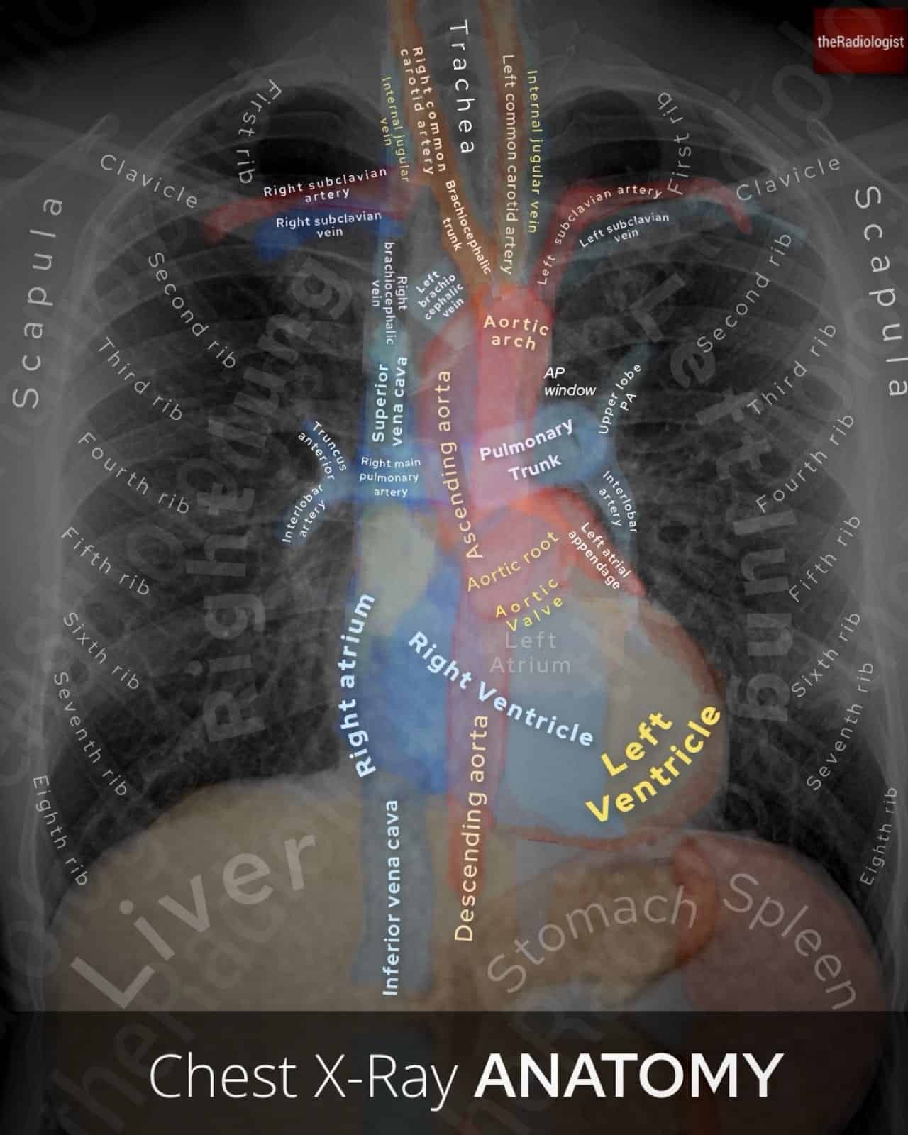
Chest X-Ray
Here we have an annotated view of a PA Chest X-ray as well as a view showing bone anatomy on a Chest X-Ray.
→ Go through the detail and learn a review system to assess these X-Rays here.
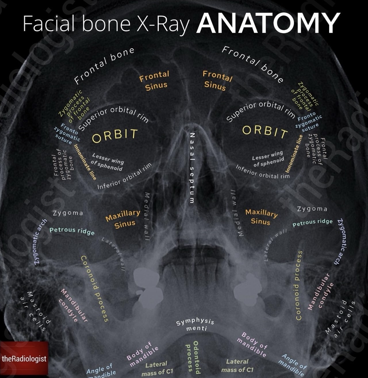
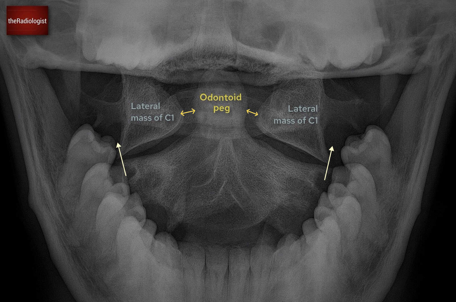
Facial bone X-Ray
Facial bone X-Rays have been superseded by CT in some parts of the world but in some places still have a role in diagnosis. Here we have an annotated view of a PA Chest X-ray as well as a view showing bone anatomy on a Chest X-Ray.
→ Go through the detail and learn a review system to assess these X-Rays here.
Spinal
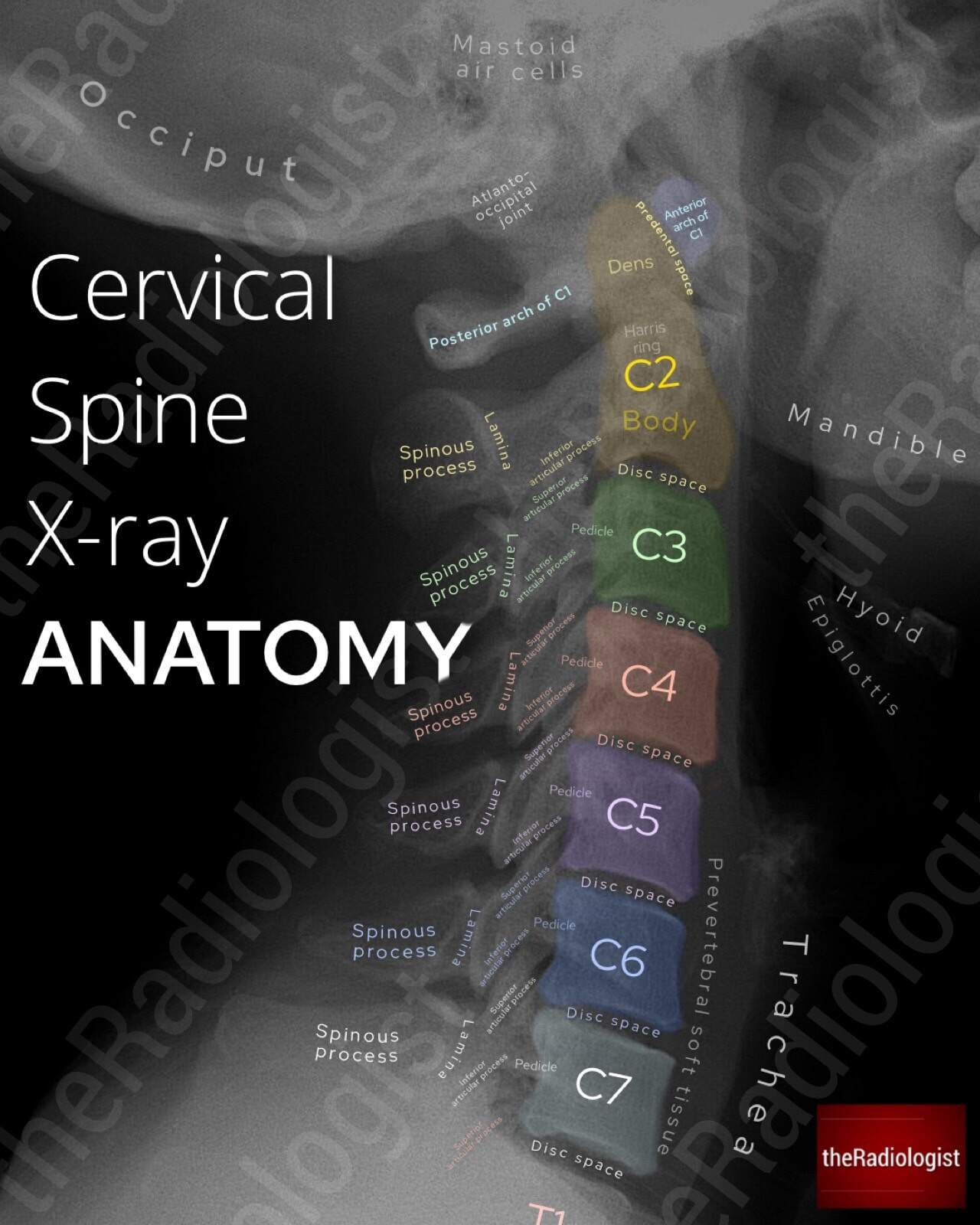
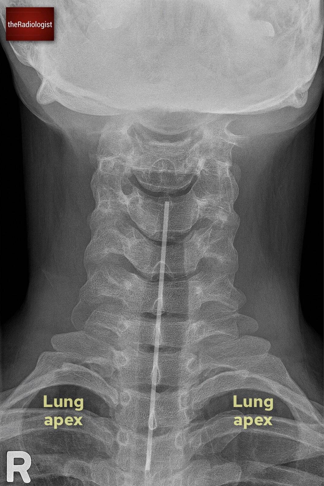
Cervical spine X-Ray
While CT is the gold standard, X-Rays still provide diagnostic use in many parts of the world. Here we have an annotated views of a lateral cervical spine X-Ray.
→ Learn a review system to help go through these X-Rays here.
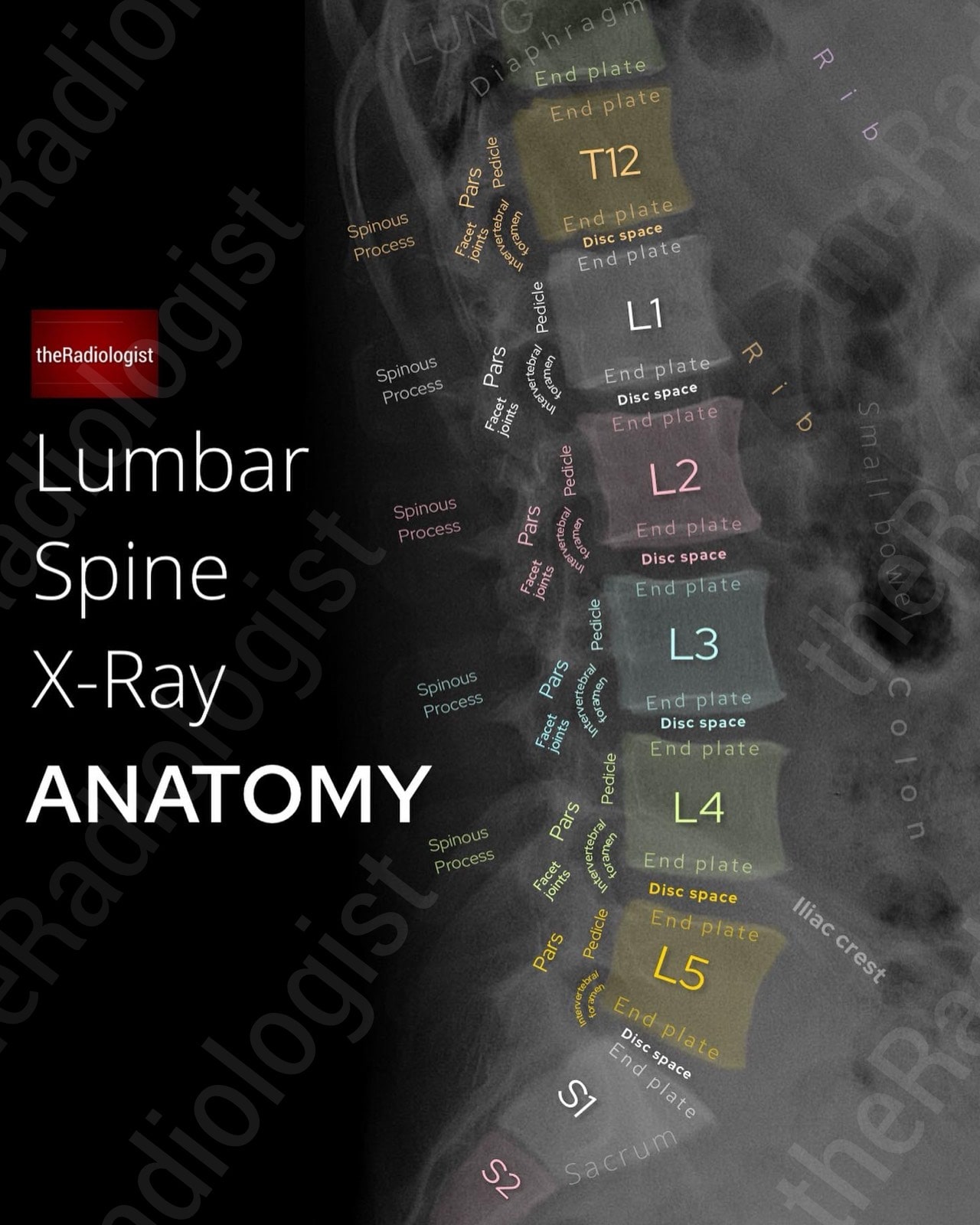
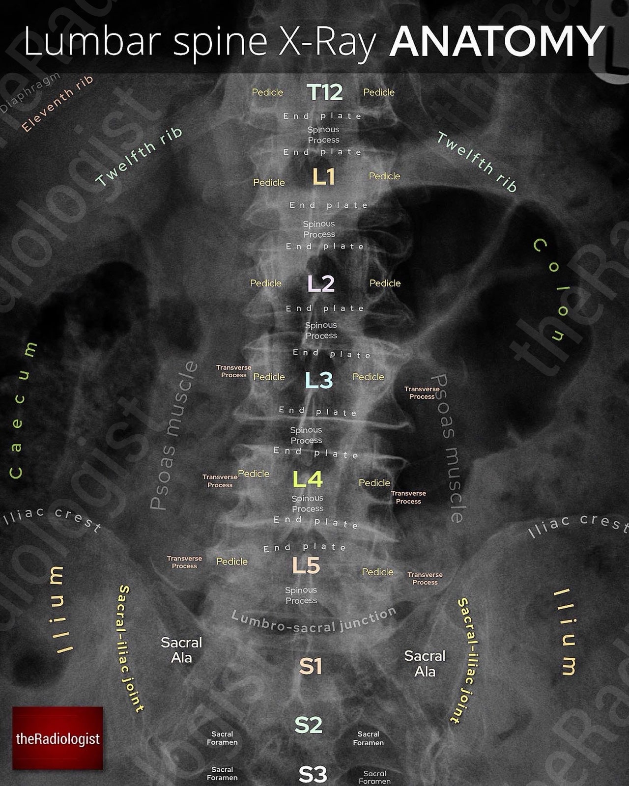
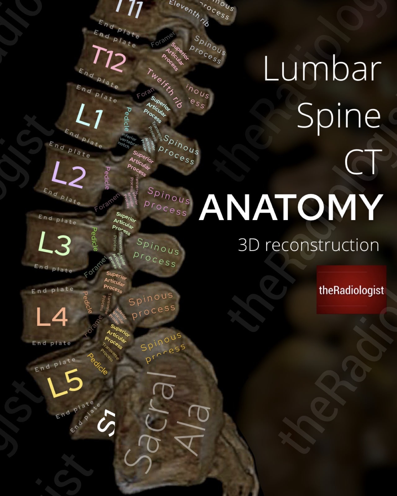
Lumbar spine X-Ray
Here we have an annotated view of a lateral lumbar spine X-Ray.
→ Go through the detail and learn a review system to assess these X-Rays here.
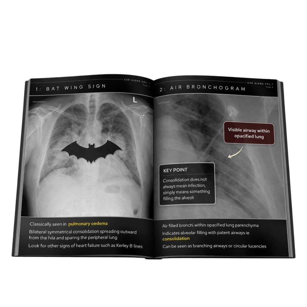
Free guide: 20 Chest X-Ray signs you need to recognise
Downloaded by 10,000+ healthcare professionals.
Get 20 annotated chest X-ray signs with clear teaching points and explanations. Written by a Consultant Radiologist, this free guide is designed to help you recognise important findings more confidently when reviewing chest X-Rays.


More to explore
You’ve got the framework now let’s put it to work. Dive into a related case to see these findings on real images, or explore another guide to build out your systematic approach. That’s where it really starts to stick.
HOW TO READ A HAND X-RAY
Hand and wrist X-Ray anatomy, views and key review areas
HOW TO READ A SHOULDER X-RAY
Shoulder X-ray anatomy, views and key alignment checks
HOW TO ASSESS ELBOW FAT PADS
How to assess elbow fat pads on a lateral elbow X-Ray