What acute appendicitis looks like on CT
This CT KUB was done for suspected renal colic, but the key finding wasn’t in the urinary tract. In this case we show how to assess the right iliac fossa for the appendix, recognise signs of appendicitis, and look for complications like perforation or abscess.
You’ll also learn how to assess for incidental findings on CT KUB, a crucial skill when the primary diagnosis isn’t what you expected.
A male in his 30s presents with right sided abdominal pain. Here is a non contrast CT KUB.
Tap below to open the interactive case. Then give it a few seconds to fully load.
Here is a video explanation of this case: click full screen in the bottom right corner to make it big. If you prefer though I go through this in the text explanation below.
Here we have a non contrast CT KUB looking for renal tract calculi. However in this case the kidneys both look normal with no hydronephrosis and no bright renal calculi. If we follow the ureters down there are no definite ureteric calculi. Although there are some right sided pelvic foci of calcification these do not definitely run in the line of the ureter and are more likely phleboliths rather than ureteric calculi.
Make sure you have a look at the bladder as well for calculi – however there are none in this case. There is however an abnormality within the right iliac fossa – always make sure to look for alternative causes of pain.
When you suspect a right iliac fossa (RIF) abnormality, first start by identifying the caecum. I do this by tracing the colon all the way from the rectum back to the origin. Once you find this, in normal circumstances you will find two things communicating with it. Firstly the terminal ileum and then the appendix.
You should be able to recognise the terminal ileum as communicating with the rest of the small bowel but also look out for some fat density which you will normally find with a normal ileocaecal valve as we can see below.
Tap play to start the video. Find the terminal ileum by identifying the distal small bowel and finding the ileocaecal valve.
When you suspect a right iliac fossa (RIF) abnormality, first start by identifying the caecum. I do this by tracing the colon all the way from the rectum back to the origin. Once you find this, in normal circumstances you will find two things communicating with it. Firstly the terminal ileum and then the appendix.
You should be able to recognise the terminal ileum as communicating with the rest of the small bowel but also look out for some fat density which you will normally find with a normal ileocaecal valve as we can see below.
Tap play to start the video. Separate to the terminal ileum we can see a second structure communicating with the caecum – this is a dilated appendix.
When we follow the appendix in this case we can see it is enlarged with surrounding fat stranding – this is acute appendicitis. It can sometimes be difficult on CT to make the call.
I usually use the diameter of the appendix and the presence of surrounding fat stranding to help me make the call. A high density appendicolith within the appendix also adds weight to the diagnosis.
What does appendicitis look like on CT?
| Diameter | Periappendiceal fat stranding | Diagnosis |
|---|---|---|
| <7 mm | – | Appendicitis not likely: consider other diagnoses |
| >7 mm | – | Indeterminate: consider appendiceal tumour if no acute pain. Look for high density appendicolith which may add weight to appendicitis. |
| >7 mm | + | Acute appendicitis |
One you find appendicitis look for complications:
Other things to think about when it comes to appendicitis:
This case shows what an inflamed appendix looks like on a CT scan, with a dilated appendix and surrounding periappendiceal inflammatory change. But did you spot the other incidental findings? Firstly there are gallstones within the gallbladder.
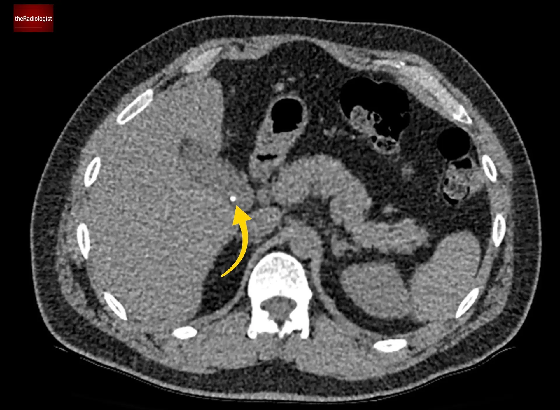
The yellow arrow shows us a gallstone within the gallbladder.
There is an umbilical hernia but most importantly there is a soft tissue lesion within the subcutaneous tissues which was worked up after the scan, resected and proven to be a soft tissue sarcoma.
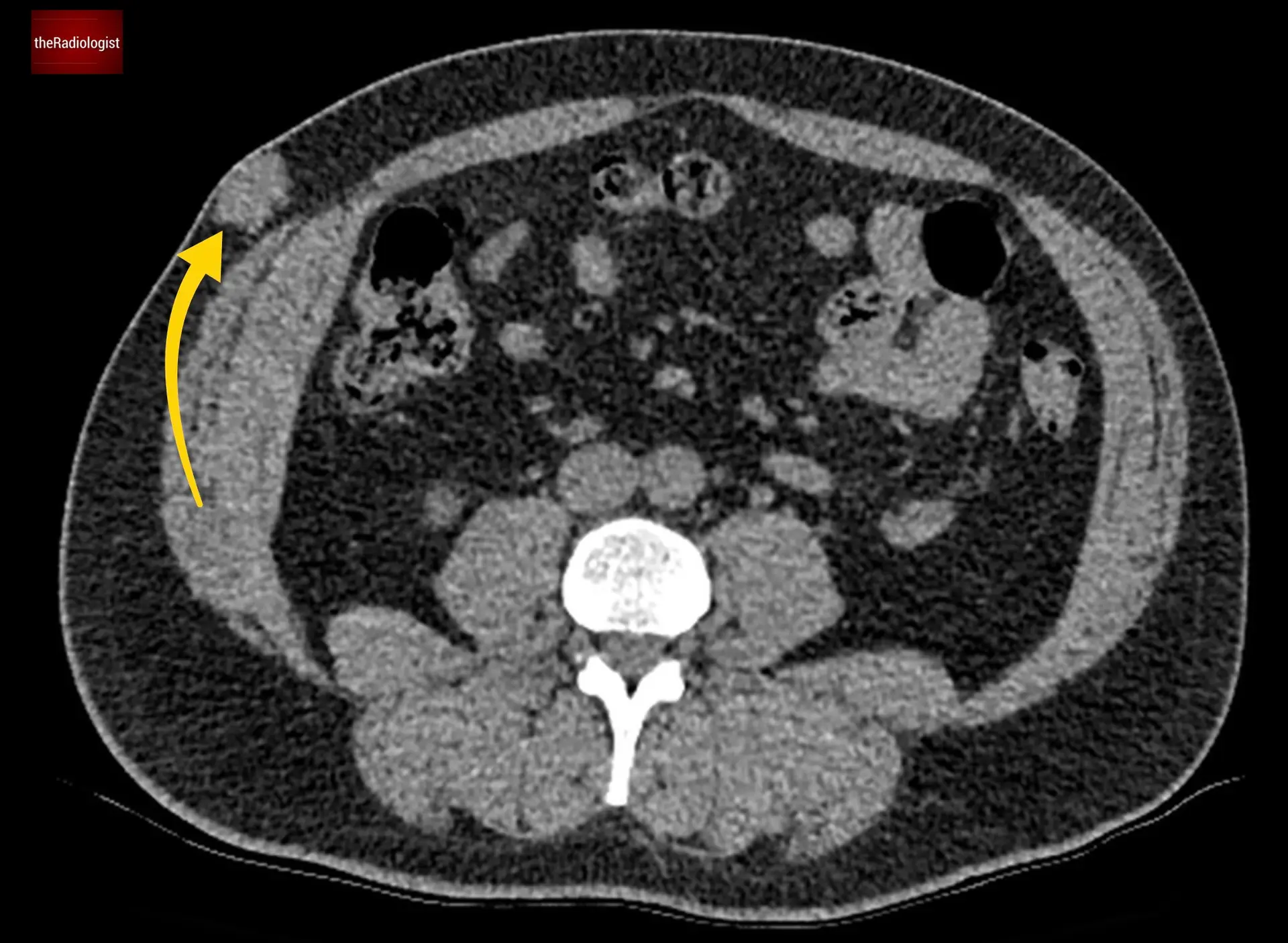
Remember to assess the peripheral soft tissues on CT: here we have an incidental lesion which later proved to be a sarcoma, thankfully resected early.
In normal circumstances you should find two structures communicating with the caecum: the terminal ileum which heads to the ileocaecal valve with its characteristic fat density as well as the appendix.
Downloaded by 10,000+ healthcare professionals.
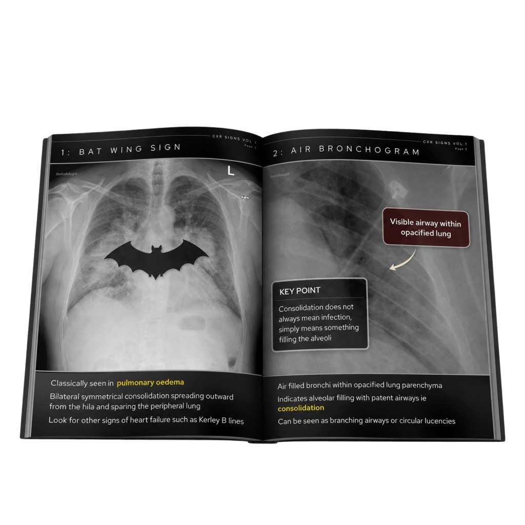
Get 20 annotated chest X-ray signs with clear teaching points and explanations. Written by a Consultant Radiologist, this free guide is designed to help you recognise important findings more confidently when reviewing chest X-Rays.
You’ve got the framework now let’s put it to work. Dive into a related case to see these findings on real images, or explore another guide to build out your systematic approach. That’s where it really starts to stick.
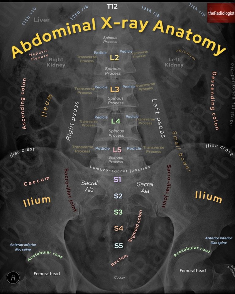
A systematic approach to bowel gas pattern, obstruction and key checks
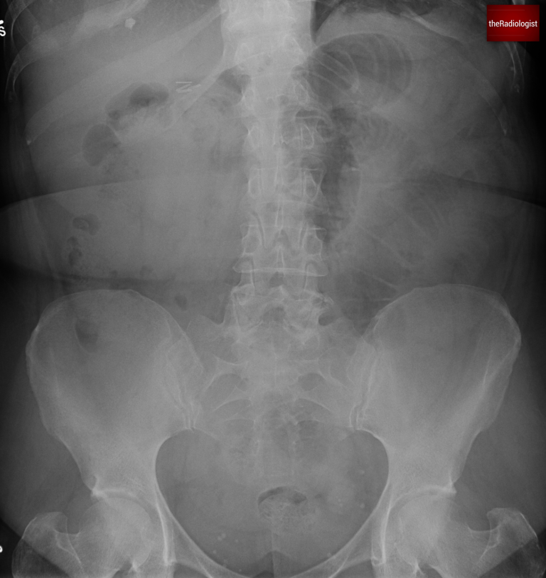
Dilated bowel loops and key review areas on CT and X-Ray
Radiology case going through the expected CT and MRI findings
We use cookies to make this website work and to understand how it is used. You can accept all cookies, use essential cookies only or manage your preferences.