Fat pads might seem like a small detail, but they can make a big difference in diagnosing elbow injuries. In this case, we look at how to assess the anterior and posterior fat pads on an elbow X-ray, even when the image isn’t perfect.
You’ll learn the key fat pad rules, why their position matters, and how they can point to an occult fracture like a subtle radial head break. A quick, high-yield guide for spotting hidden trauma on elbow films.
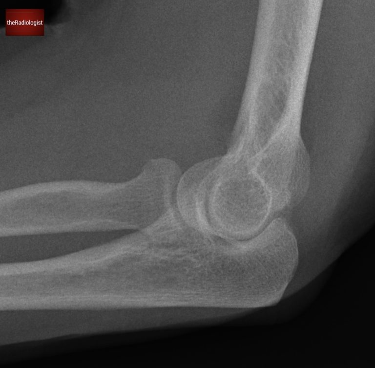
Have a look at this case of elbow trauma. The projection is suboptimal but regardless, there’s a clear abnormality. The key to interpreting this film lies in recognizing the significance of fat pads. Let’s break it down.
Lateral Elbow X-Ray
Here is a video explanation of this case: click full screen in the bottom right corner to make it big. If you prefer though I go through this in the text explanation below.
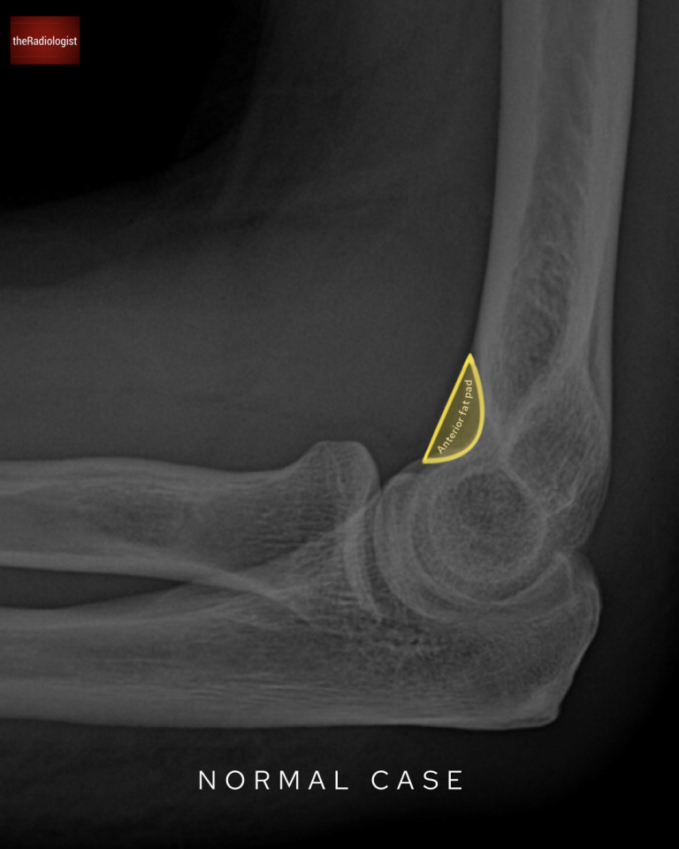
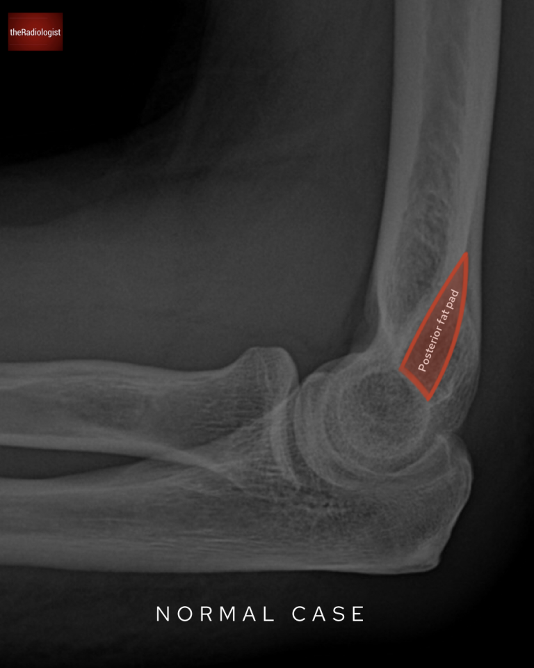
To understand what’s abnormal, let’s first review normal fat pad anatomy:
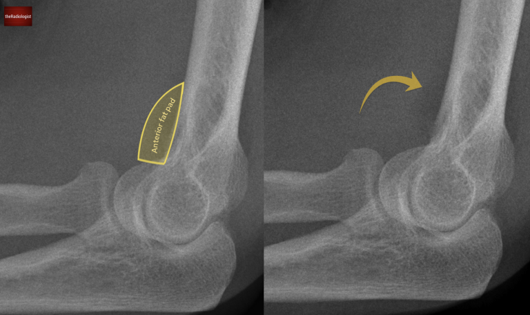
Anterior fat pad on a normal elbow X-Ray.
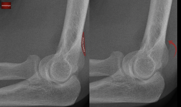
Posterior fat pad on a normal elbow X-Ray: this is normally obscured.
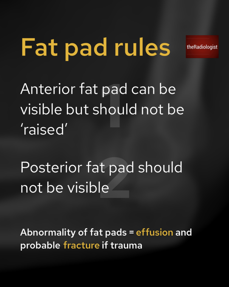
There are two essential rules to remember when assessing fat pads on an elbow X-ray:
The fat pad rules.
Going back to our case, the alignment isn’t the best on our X-Ray in that the capitellum and trochlea do not overlap each other. However sometimes we have to deal with suboptimal images and still apply the rules we know. Let’s apply our fat pad rules:
Instead of a thin stripe the anterior fat pad is clearly raised, signaling something abnormal. Now let’s have a look for a posterior fat pad.
A ‘raised’ abnormal anterior fat pad can be seen.
The posterior fat pad is visible in this case when in normal circumstances it should be obscured.
What does this mean? The presence of abnormal fat pads indicates an elbow joint effusion. In the context of trauma, this strongly suggests an intra-articular fracture.
We can see a posterior fat pad which in normal circumstances should be obscured like the case above.
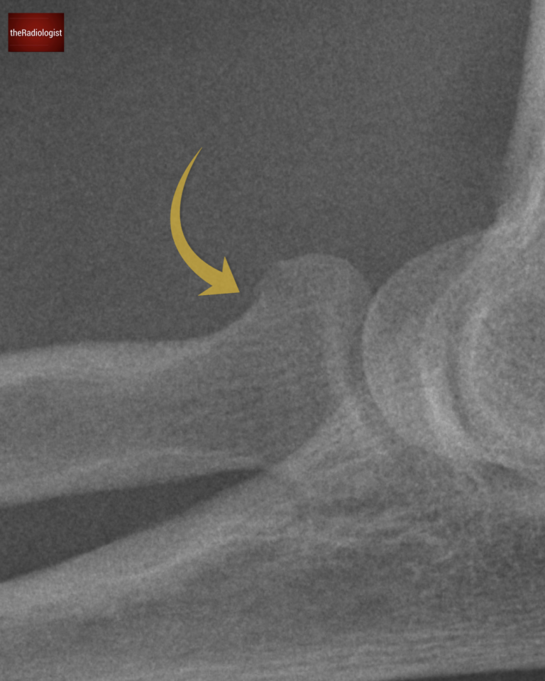
In adults, the most common elbow fracture is a radial head fracture, so that’s the first place to check.
Looking closely we can see a subtle fracture at the radial head.
Don’t forget the fat pad rules!
The anterior fat pad can be visible but not raised.
The posterior fat pad should never be visible.
Downloaded by 10,000+ healthcare professionals.
Get 20 annotated chest X-ray signs with clear teaching points and explanations. Written by a Consultant Radiologist, this free guide is designed to help you recognise important findings more confidently when reviewing chest X-Rays.
You’ve got the framework now let’s put it to work. Dive into a related case to see these findings on real images, or explore another guide to build out your systematic approach. That’s where it really starts to stick.
AP, lateral and labelled anatomy for interpretation
Learn about the X-Ray features, anatomy and treatment
Anterior dislocation signs, key views and associated fractures
We use cookies to make this website work and to understand how it is used. You can accept all cookies, use essential cookies only or manage your preferences.