A man in his 40s presents with finger pain and swelling after a hyperextension injury. The X-ray looks normal at first glance, but there’s more to see.
In this case, we’ll walk through the importance of two views in finger trauma, show how to spot a volar plate injury, and explain why the lateral X-ray is key to making the diagnosis.
A male in his 40s presents with pain and swelling in his index finger following a hyperextension injury. Have a look at the X-Ray below.
PA (left) and lateral (right) views of the index finger
Video explanation
Here is a video explanation of this case: click full screen in the bottom right corner to make it big. If you prefer though I go through this in the text explanation below.
Assessing the PA view
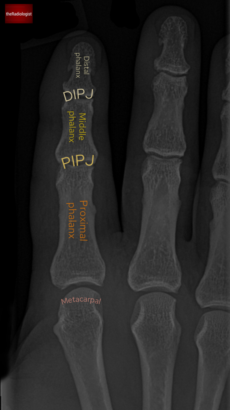
Let’s begin with the frontal X-ray of the index finger.
Bones: Start by identifying the metacarpal, proximal phalanx, middle phalanx, and distal phalanx.
Soft Tissue Swelling: Comparing the index finger to the middle finger, there’s visible soft tissue swelling, a hallmark sign of trauma.
Alignment: Each phalanx lines up well with the adjacent one, so the alignment appears normal.
Fracture Search:
Look for breaks in the cortex or lucent lines, the key signs of a fracture.
Zoom in to examine the cortex of each bone closely.
On this view, no obvious fracture is seen.
Annotated image of the PA view of the X-Ray showing the basic anatomy.
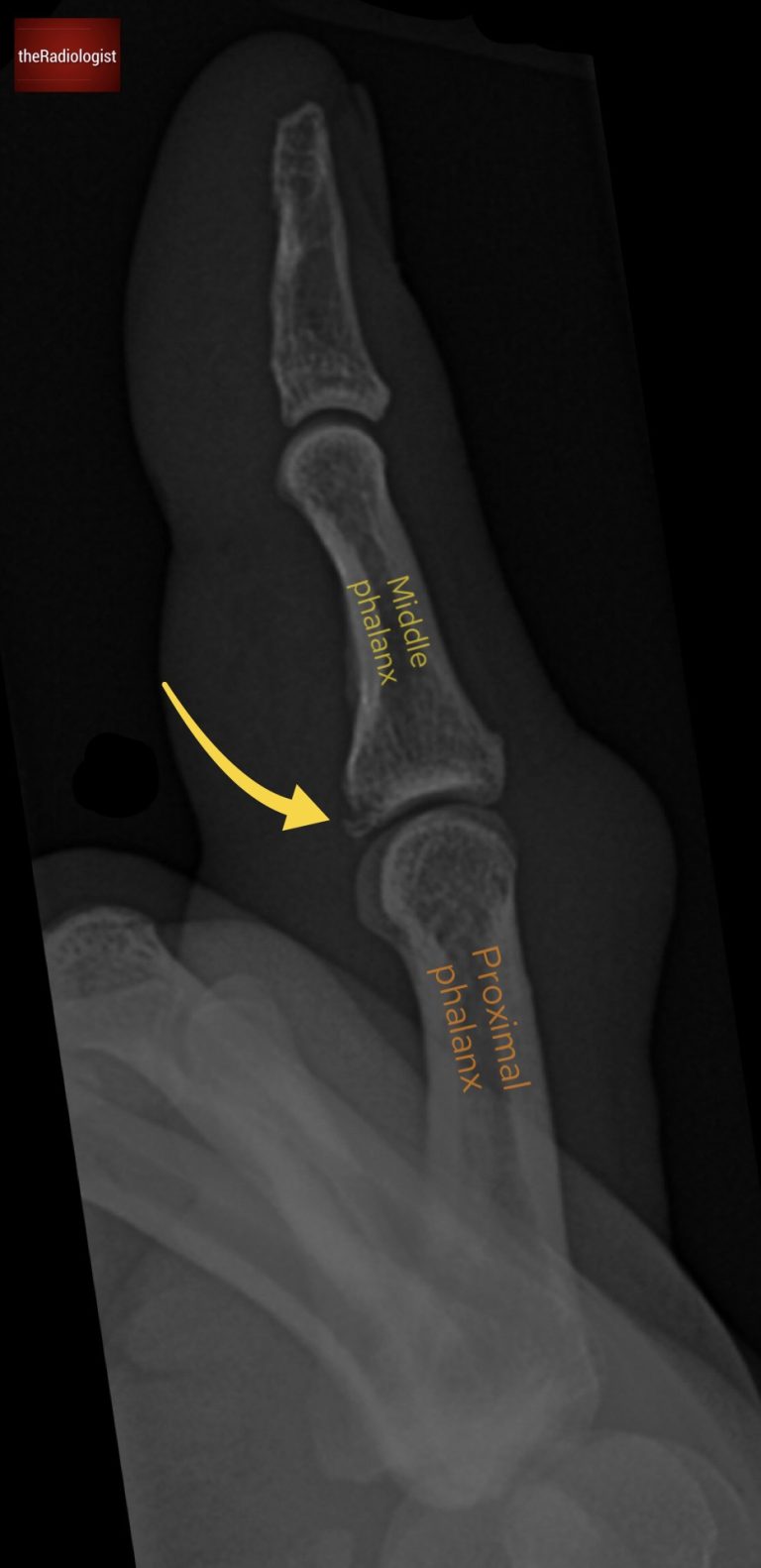
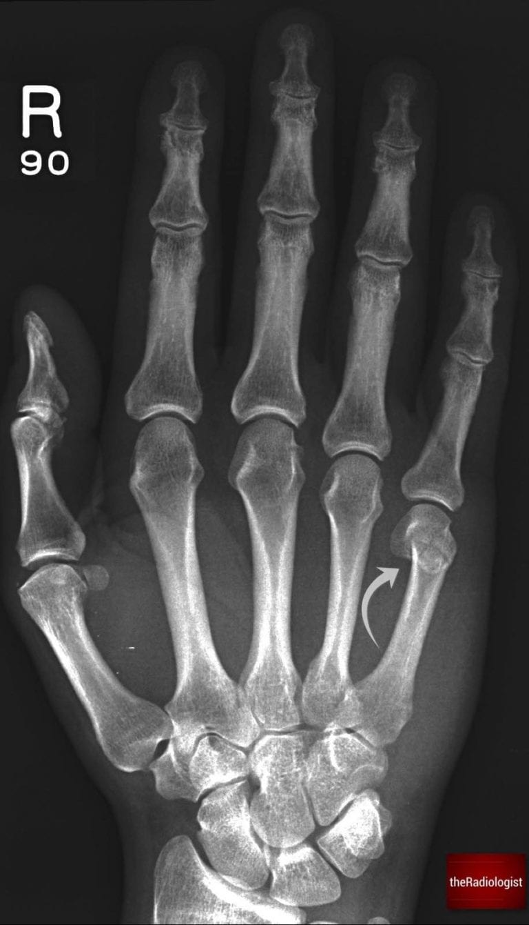
Assessing the lateral view
In trauma imaging, one view is never enough. A single projection may miss critical findings. Let’s move to the lateral X-ray to get a clearer picture.
On the lateral X-ray:
Soft Tissue Swelling: The swelling seen on the frontal view is confirmed here.
Fracture Identification: This time, a break in the cortex is visible at the base of the middle phalanx, just above the proximal interphalangeal (PIP) joint.
This location is classic for a volar plate injury.
Lateral view shows a fracture at the base of the middle phalanx.
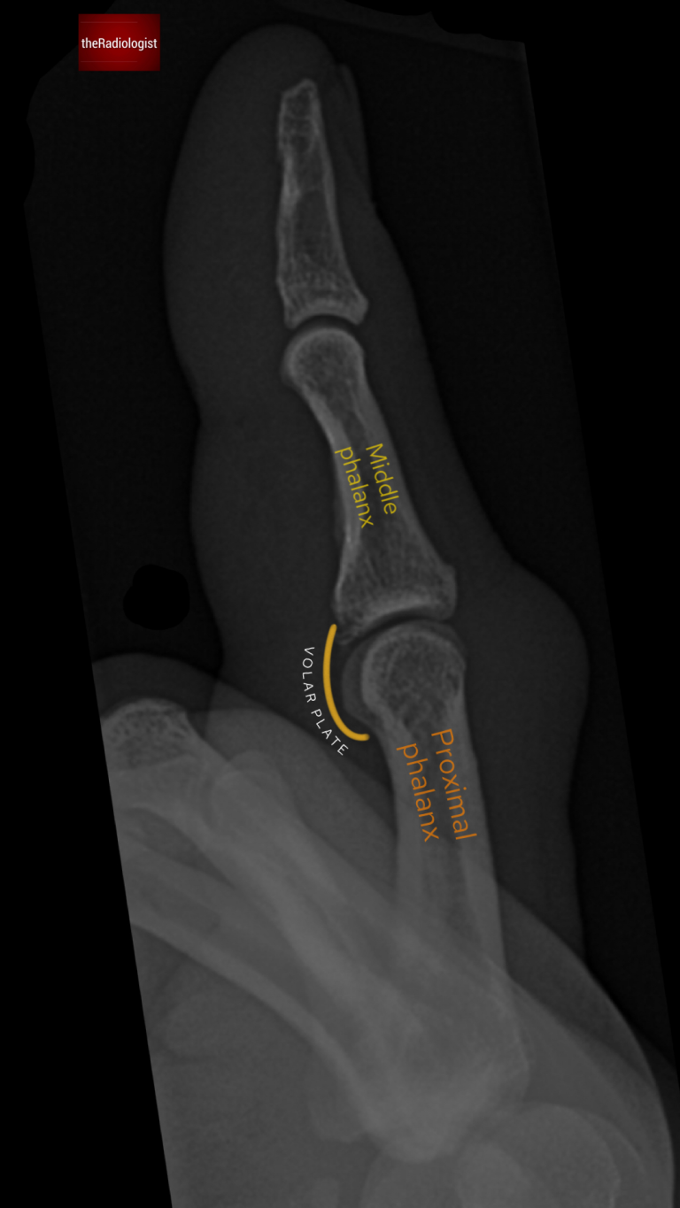
The volar plate
The volar plate is a fibrocartilaginous structure that stabilizes the PIP joint and prevents hyperextension.
It originates from the proximal phalanx and inserts onto the base of the middle phalanx.
Hyperextension can cause partial or complete rupture, often leading to an avulsion fracture at the middle phalanx base.
Location of Rupture
Most commonly at the base of the middle phalanx due to checkrein ligaments reinforcing the proximal phalanx insertion.
Fracture Pattern
The injury may present with a visible avulsion fracture fragment. In severe cases, there can be subluxation or even dislocation of the PIP joint.
The volar plate stablises the proximal interphalangeal joint (PIPJ) and inserts onto the base of the middle phalanx.
Treatment
Most volar plate injuries are managed conservatively with splinting.
When should we intervene?
Surgical intervention is considered if the fracture involves more than 40% of the joint surface or if it cannot be reduced to less than 30 degrees of flexion.
KEY POINT
In hyperextension injuries remember to interrogate the middle phalanx on the lateral view so as to not miss a volar plate injury.
FREE GUIDE
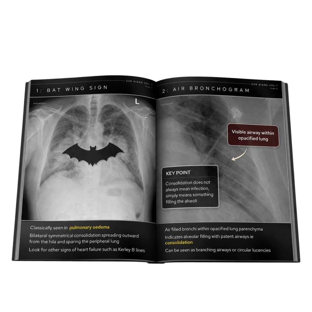
Free guide: 20 Chest X-Ray signs you need to recognise
Downloaded by 10,000+ healthcare professionals. Get 20 annotated chest X-ray signs with clear teaching points and explanations. Written by a Consultant Radiologist, this free guide is designed to help you recognise important findings more confidently when reviewing chest X-Rays.
You’ve got the framework now let’s put it to work. Dive into a related case to see these findings on real images, or explore another guide to build out your systematic approach. That’s where it really starts to stick.