A woman in her 40s presents after a head injury and loss of consciousness. A non-contrast CT is performed, and a high-density lesion is spotted in the occipital lobe. But the underlying diagnosis is not clear.
This case walks you through how to assess an unusual hyperdense lesion on CT, when to suspect something beyond trauma, and how MRI can confirm the diagnosis.
A useful reminder that not every bright spot on a head CT is a bleed.
A female patient in her 40s presented to the emergency department after a head injury. She had lost consciousness earlier the same day and underwent a non-contrast CT scan of her head.
Have a look at the scan below.
I know you want to get going but you may need to wait a few seconds for the scan to load. Tap the first icon on the left to scroll.
Here is a video explanation of this case: click full screen in the bottom right corner to make it big. If you prefer though I go through this in the text explanation below.
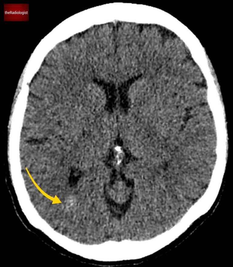
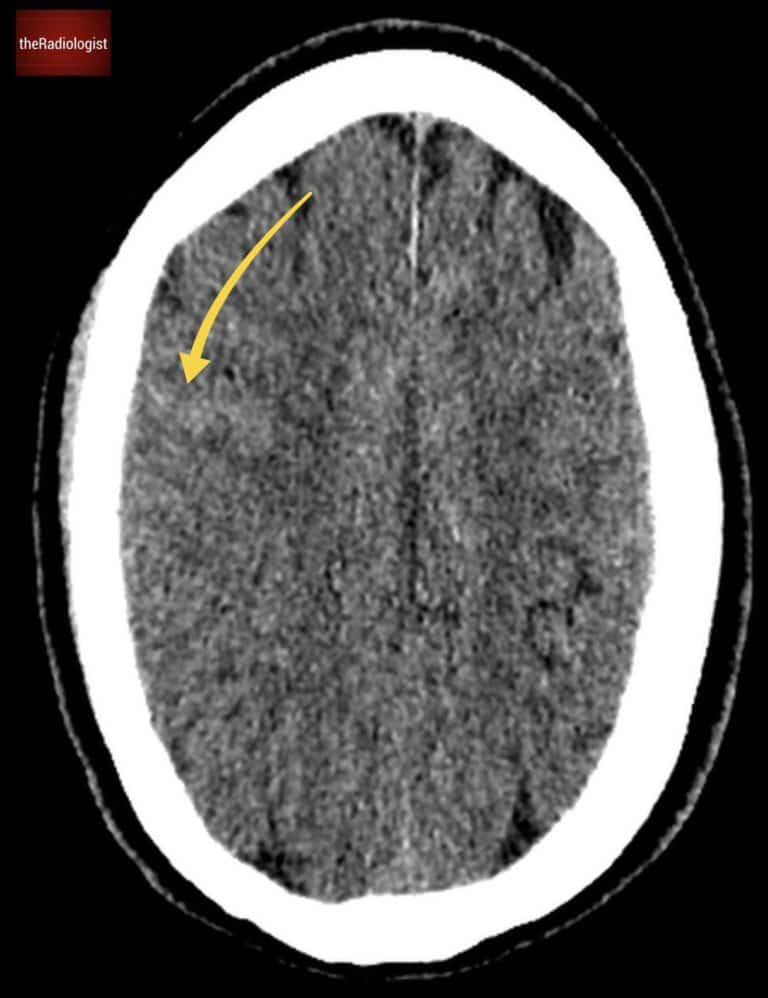
Looking at the CT scan, on first inspection there is a high density focus within the right occipital lobe. Now any high density on an acute CT head warrants suspicion for acute haemorrhage.
On closer inspection the lesion appears heterogeneous with possible flecks of calcification. There is no mass effect so the surrounding structures such as the right lateral ventricle are not distorted. Also there is no oedema (a low density rim would suggest vasogenic oedema).
Did you spot the high density lesion within the right occipital lobe?
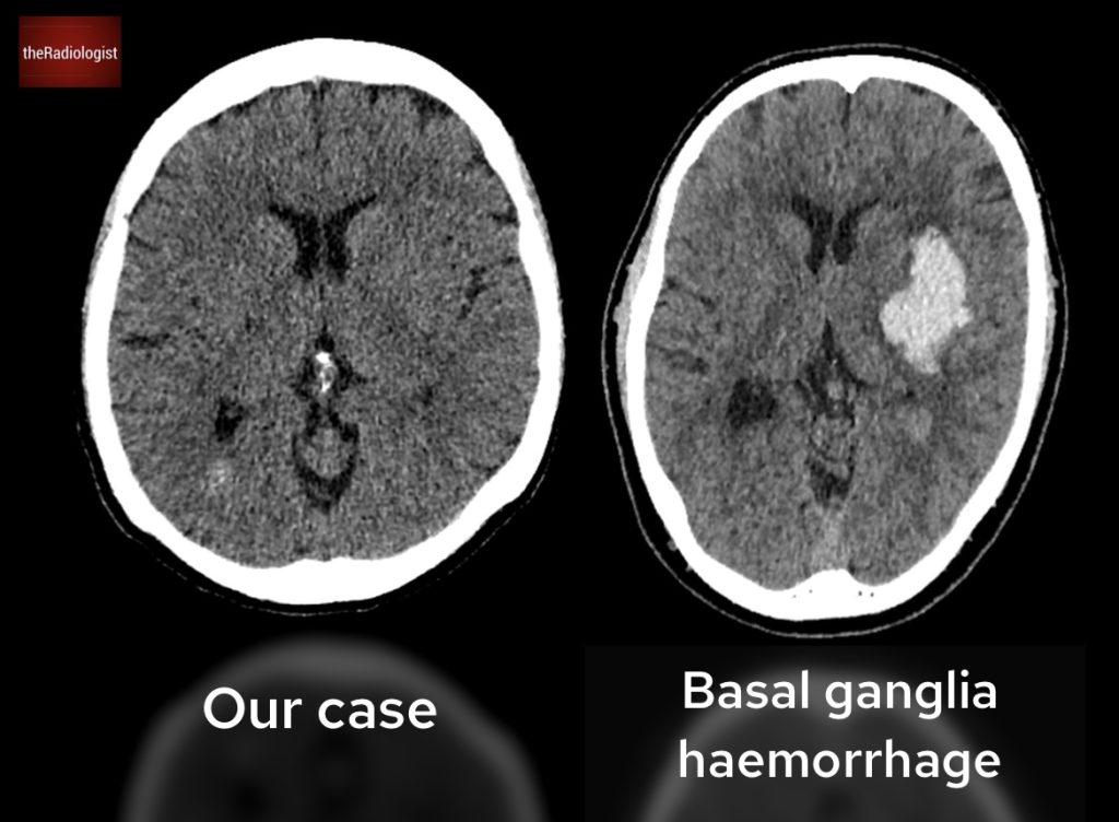
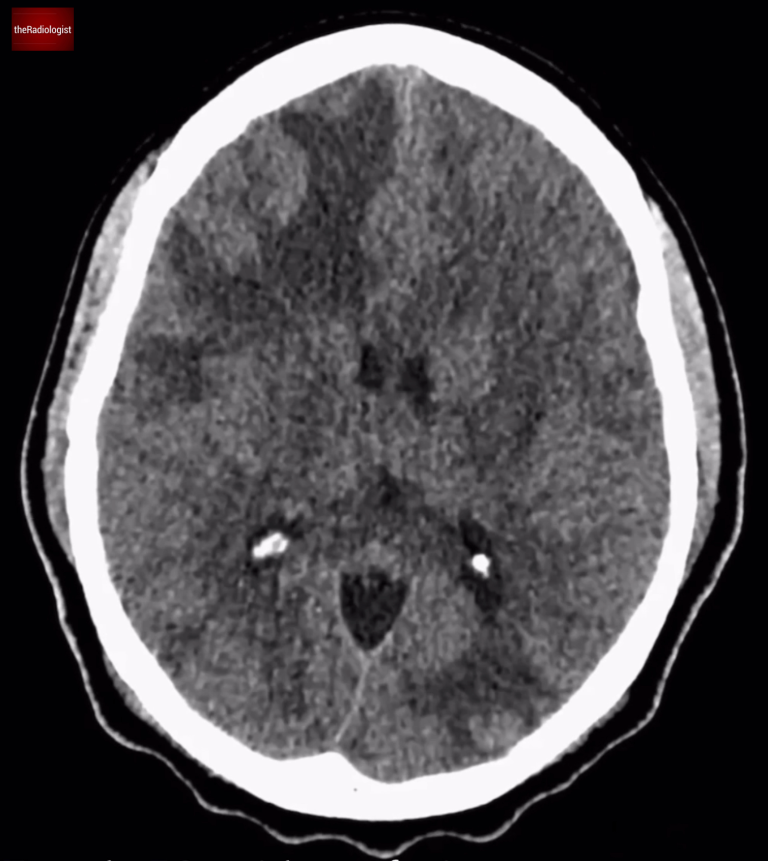
Now, let’s compare this to a known case of intraparenchymal haemorrhage:
Firstly the lesion in our case is not as dense as a typical acute haemorrhage.
There’s no surrounding oedema.
The presence of calcification further raises suspicion for something other than trauma-related haemorrhage.
Compare our case (left) with a case of confirmed basal ganglia haemorrhage on the right.
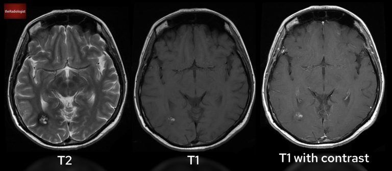
Given the unusual CT appearance, the patient underwent an MRI. Here’s what the MRI revealed:
Have a look at T2 (left), T1 (centre) and T1 post gadolinium (right) sequences.
The imaging findings point to a cavernous malformation (also known as a cavernoma or cerebral cavernous venous malformation).
What Are Cavernous Malformations?
If found incidentally, cavernous malformations are often managed conservatively and monitored.
Surgical resection may be considered in cases of:
When evaluating a possible intracerebral bleed on CT that lacks the classic brightness of a cerebral haemorrhage think about a cavernous malformation.
These lesions are best confirmed with MRI, where their characteristic features usually show through.
Downloaded by 10,000+ healthcare professionals.
Get 20 annotated chest X-ray signs with clear teaching points and explanations. Written by a Consultant Radiologist, this free guide is designed to help you recognise important findings more confidently when reviewing chest X-Rays.
You’ve got the framework now let’s put it to work. Dive into a related case to see these findings on real images, or explore another guide to build out your systematic approach. That’s where it really starts to stick.
CT head and CT angiogram features, review areas and pitfalls
Review the anatomy, early signs of an MCA infarct and CT angiogram findings
Brain CT and MRI features, mass effect and differential diagnosis
We use cookies to make this website work and to understand how it is used. You can accept all cookies, use essential cookies only or manage your preferences.