Radiology case with CXR and CT chest features
Septic emboli can be easy to miss but dangerous to overlook. Recognising the imaging features and knowing when to suspect them is key, especially in young patients with systemic symptoms and there are some classic imaging findings.
In this case, we walk through the chest X-ray and CT findings, explore the differential for multiple lung nodules, and highlight the steps to track down the source. A useful case to help you spot the clues, think through the differential, and see how imaging can steer the whole diagnosis.
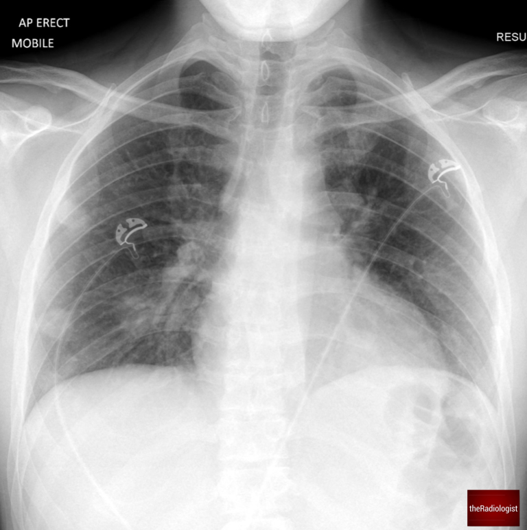
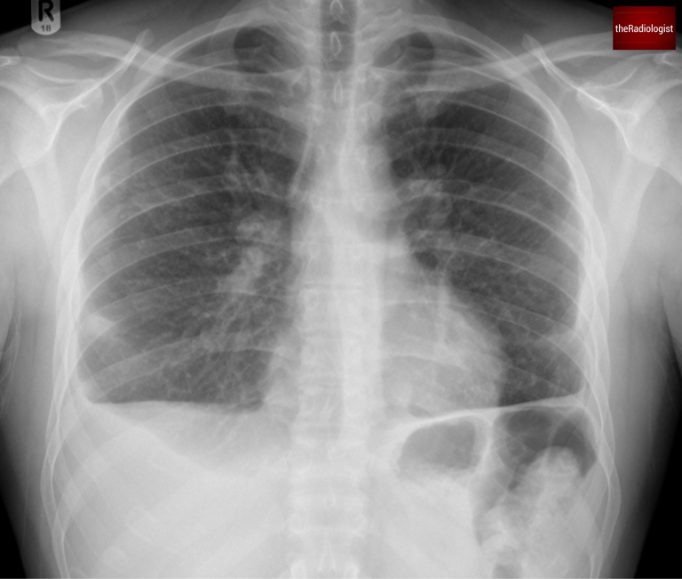
A male in his 20s presents to the emergency department with fever, cough, diarrhoea, and abdominal pain. An anterior-posterior (AP) chest X-ray was performed in ED – we already know by the fact this is AP that the patient is unlikely to be unwell.
AP Chest X-Ray of a male in his 20s
Here is a video explanation of this case: click full screen in the bottom right corner to make it big. If you prefer though I go through this in the text explanation below.
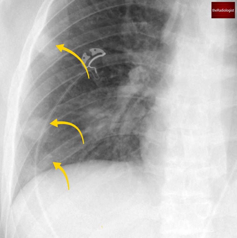
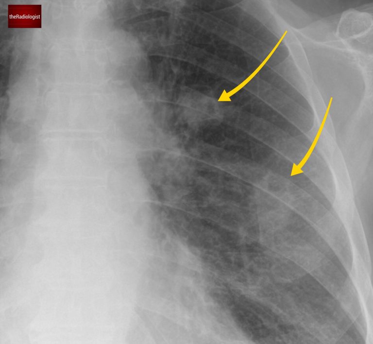
The chest X-ray shows several nodules in the right lung, predominantly in the middle and lower zones.
Note that these nodules are notably peripheral, with no evidence of pleural effusion.
There are several peripheral nodules within the right lung (yellow arrow). There are further nodules seen centrally overlying the hilar vessels in addtion.
In young, acutely ill patients, the differential for multiple lung nodules includes:
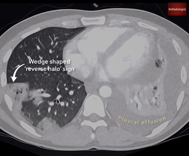
A subsequent CT scan clarified the findings:
The reverse halo sign is seen in various conditions but is commonly associated with lung infarction, as seen in septic emboli.
At this stage, septic emboli became the leading differential diagnosis. However, identifying the source of infection was critical to confirm and guide treatment.
There is a wedge shaped region of consolidation and groundglass opacity on the right and a pleural effusion on the left.
Septic emboli originate from infections that spread via the bloodstream. Common sources include:
In particular right-sided endocarditis linked to intravenous drug use. Check for heart murmurs and/or perform an echocardiogram.
Consider indwelling catheters, soft tissue infections and intravenous drug use – examine the groin both physically and on CT.
Conditions like tonsillitis or pharyngitis can spread to the bloodstream.
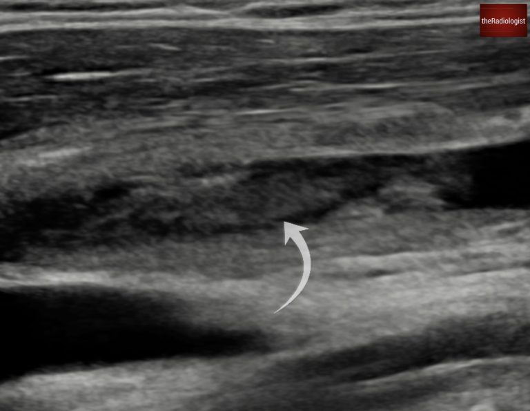
In this case, the patient reported a sore throat one week prior to becoming acutely ill. An ultrasound of the internal jugular vein was performed, revealing a large clot, confirming Lemierre Syndrome.
There is thrombus within the internal jugular vein.
Lemierre syndrome is a condition where an infection (commonly bacterial) in the head or neck, such as pharyngitis or tonsillitis, causes thrombophlebitis of the internal jugular vein. Fusobacterium necrophorum (a Gram negative bacillus) is responsible for the majority of infections.
This can result in septicaemia and septic emboli within the lungs and large joints, and can result in in high mortality if untreated. Treatment strategy usually involves intravenous antibiotics and anticoagulation.
The patient’s left pleural effusion was tapped and confirmed to be an empyema, a known complication of septic emboli. Radiologists inserted a chest drain to manage this.
After several weeks of treatment, including chest drains, the patient improved. A follow-up chest X-ray showed almost complete resolution of the lung opacities and pleural effusions.
Interestingly, this patient’s initial presentation included abdominal pain and diarrheoa. While unusual, a small subset of septic emboli cases can present with gastrointestinal symptoms. An abdominal CT performed at the time of diagnosis showed no abnormalities, emphasising the need to correlate symptoms with systemic infection.
A follow up chest X-Ray shows almost complete resolution of the right lung nodules.
Don’t miss septic emboli!
Peripheral cavitating nodules and wedge shaped opacities should make you think about septic emboli.
Once you see these hunt for a source: consider head and neck infection, endocarditis, soft tissue infections and indwelling devices.
Downloaded by 10,000+ healthcare professionals.
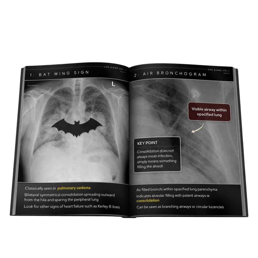
Get 20 annotated chest X-ray signs with clear teaching points and explanations. Written by a Consultant Radiologist, this free guide is designed to help you recognise important findings more confidently when reviewing chest X-Rays.
You’ve got the framework now let’s put it to work. Dive into a related case to see these findings on real images, or explore another guide to build out your systematic approach. That’s where it really starts to stick.
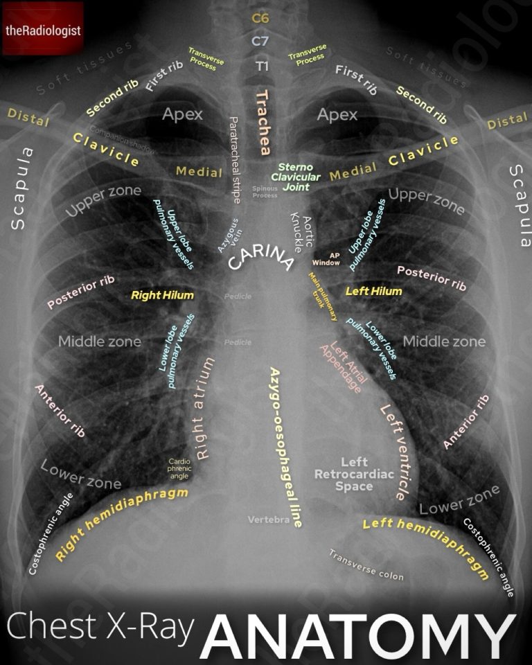
A systematic approach to anatomy, lines and tubes and review areas
How to differentiate between malignant and benign causes
Classic CXR and CT chest features and key differential
We use cookies to make this website work and to understand how it is used. You can accept all cookies, use essential cookies only or manage your preferences.