Chest X-ray and CT differential diagnosis
A man in his 80s presents unwell and his chest X-ray shows multiple lung lesions. When you see cavitation on imaging, especially when it’s widespread, the key is having a structured approach.
In this case, we break down how to recognise cavitation, work through the differential using the STARE mnemonic, and explore how to narrow things down with clinical context, CT findings, and biopsy.
A practical guide to tackling one of the more complex patterns in chest imaging.
When you’re faced with multiple cavitating lung lesions on imaging, it’s really important to approach the case methodically, as the differential diagnosis is broad – however it is manageable with a structured approach.
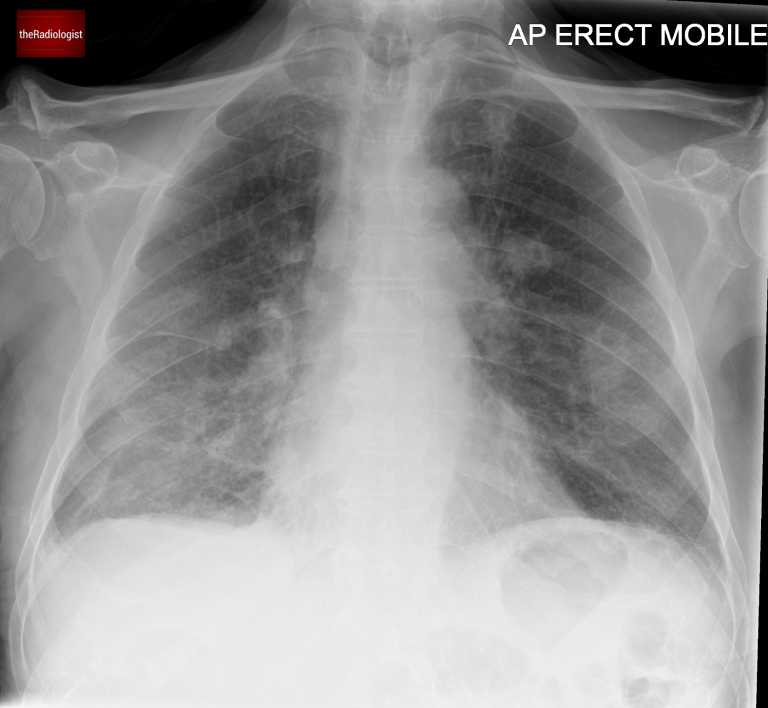
Let’s go through a case of a male in his 80s who presented unwell to the Emergency Department and had a chest X-Ray.
AP Chest X-Ray
Here is a video explanation of this case: click full screen in the bottom right corner to make it big. If you prefer though I go through this in the text explanation below.
Here we have a non contrast CT KUB looking for renal tract calculi.
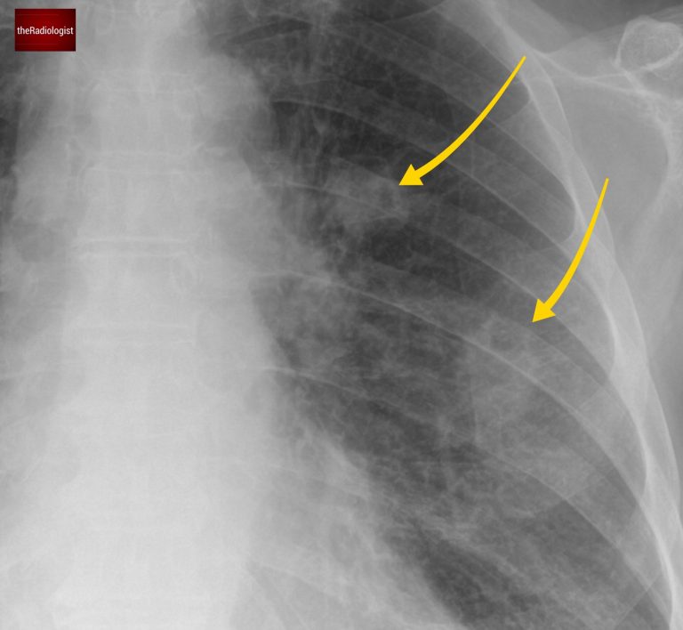
When we zoom in, we can see a bilateral abnormality. On closer inspection, some of these lesions have a small associated lucency, representing gas within the lesion.
This finding is called cavitation, and identifying it helps significantly narrow down the differential diagnosis. The differential changes as to whether we are dealing with either a single or multiple cavitating lesions. Let’s have a look at both below
Look closely and you will see multiple lung nodules, some of which show internal lucency, ie cavitation
The differential for a single cavitating lesion is usually limited:
Now, multiple cavitating lesions, like in this case, expand the differential. I like to use the mnemonic STARE:
| Cause | |
|---|---|
| S | SCC metastases |
| T | TB and atypical infection |
| A | ANCA positive vasculitis |
| R | Rheumatoid nodules |
| E | septic Emboli |
Let’s have a look at each of these in a bit more detail:
There are several causes of cavitating lung metastases but they most commonly derive from a squamous cell carcinoma, often originating from the lung or head and neck.
Always an important consideration, consider fungal infection such as aspergillosis. With TB look for involvement of the right upper lobe and apical right lower lobe, tree-in-bud nodularity and necrotic lymph nodes.
Most notably granulomatosis with polyangiitis (GPA), formerly known as Wegener’s granulomatosis.
Screening for ANCA antibodies is typically advised in these cases: I have seen several cases picked up over the years after recommending an ANCA screen.
These are usually small and peripheral, some of which can cavitate.
Also typically small and peripheral. Also look out for wedge shaped opacities – see this case for more on septic emboli.
Let’s go back to our case. On closer inspection, the X-ray reveals an abnormal aortopulmonary (AP) window.
Looking at the mediastinum there is filling of the AP window suggesting lymph node enlargement.
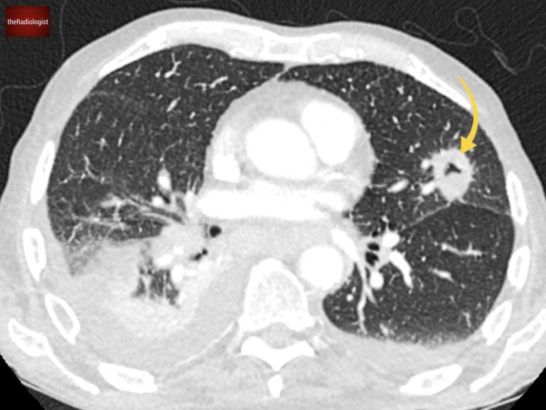
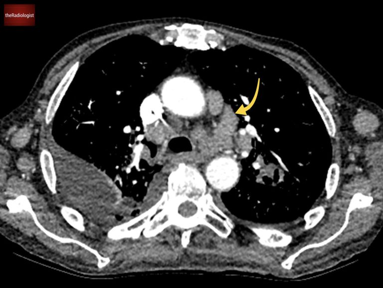
A post contrast CT scan was performed which confirmed the chest X-Ray findings of multiple cavitating lung lesions, enlarged mediastinal lymph nodes involving the aortopulmonary window as well as a bilateral pleural effusions.
The cavitating lesions are confirmed on CT.
On soft tissue windows we can see a right pleural effusion, left axillary node and enlarged mediastinal nodes. AP window nodes (arrow) are seen accounting for the X-Ray abnormality.
The diagnosis in this case came from biopsying groin lymph nodes, which revealed a metastatic neuroendocrine tumor.
This isn’t the most common cause of cavitating lung lesions, but it emphasises the need for a tissue diagnosis when imaging findings aren’t conclusive.
Use the STARE mnemonic when you see multiple cavitating lesions on X-Ray: this will cover the majority of causes and gives you a systematic approach.
Downloaded by 10,000+ healthcare professionals.
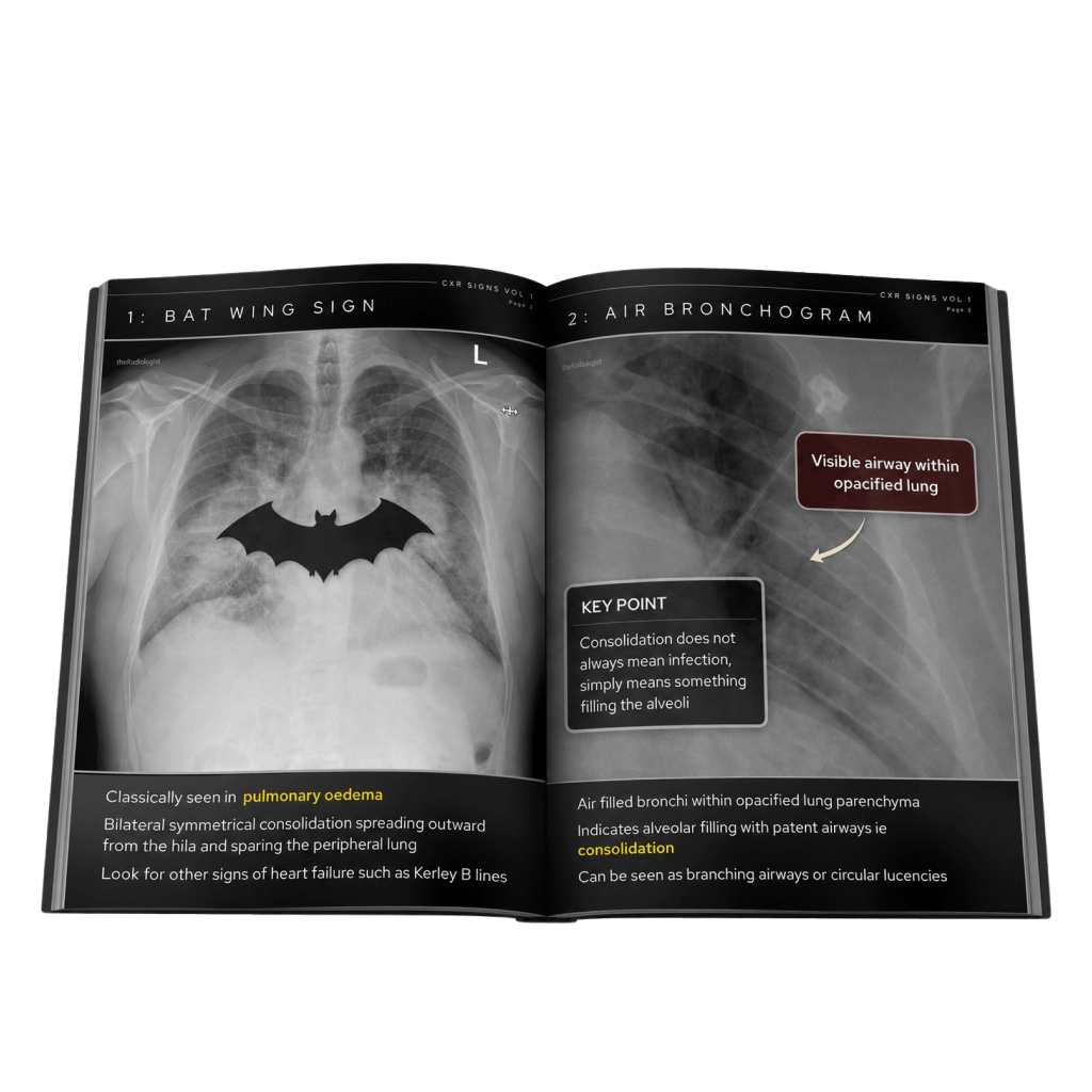
Get 20 annotated chest X-ray signs with clear teaching points and explanations. Written by a Consultant Radiologist, this free guide is designed to help you recognise important findings more confidently when reviewing chest X-Rays.
You’ve got the framework now let’s put it to work. Dive into a related case to see these findings on real images, or explore another guide to build out your systematic approach. That’s where it really starts to stick.
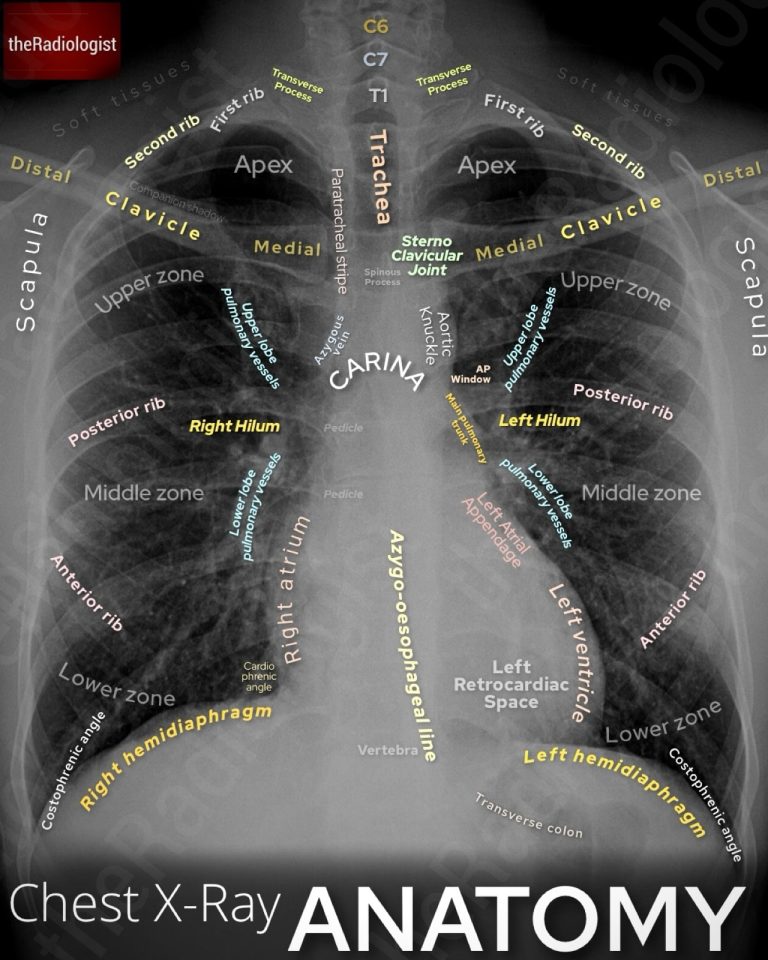
A systematic approach to anatomy, lines and tubes and review areas
How to differentiate between malignant and benign causes
Go through a case showing classic CXR and CT chest features
We use cookies to make this website work and to understand how it is used. You can accept all cookies, use essential cookies only or manage your preferences.