X-ray signs, bowel dilatation and CT features
A woman in her 60s presents with lower abdominal pain and a background of constipation. Her CT scan shows a large bowel obstruction that needs urgent attention.
In this case, we review the CT and X-ray findings, go over how to distinguish small from large bowel obstruction, explore possible causes, and discuss next steps in management.
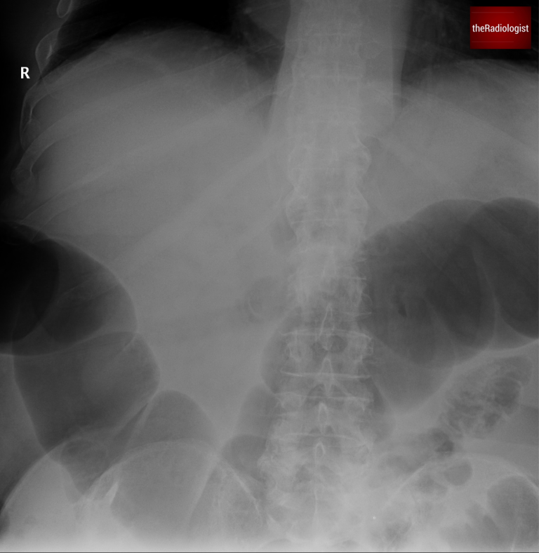
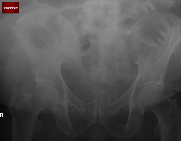
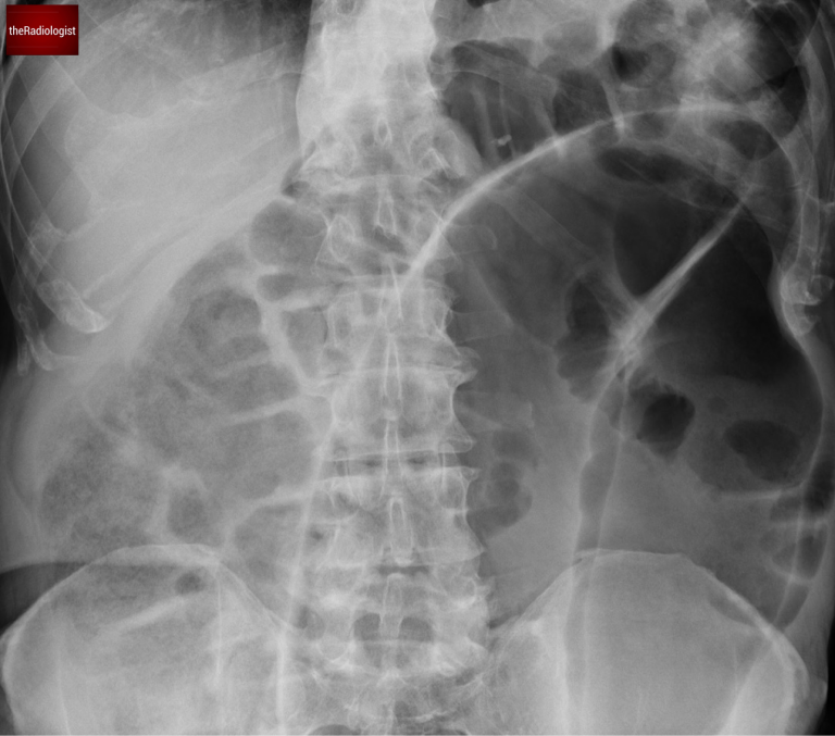
A female in her 60s presents with lower abdominal pain and absolute constipation. Here is her abdominal X-Ray (two separate images):
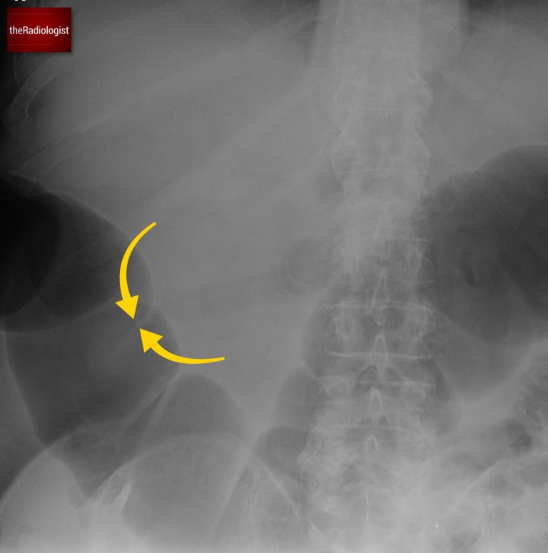
Abdominal X-Ray on presentation: image 1
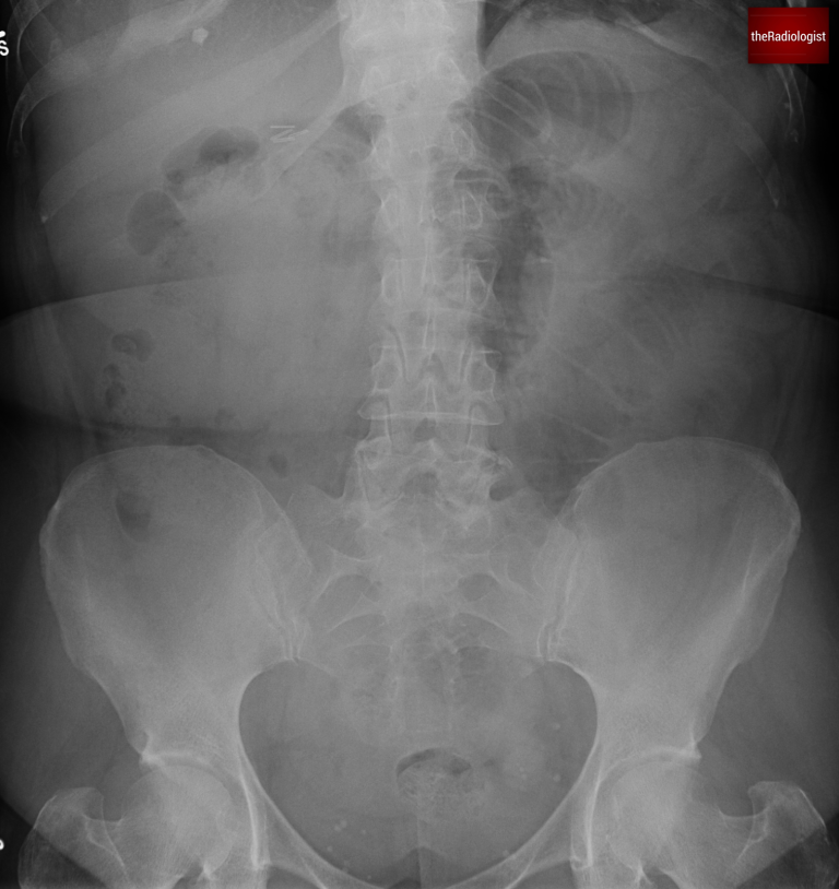
Abdominal X-Ray on presentation: image 2
Now have a look through the CT of the abdomen and pelvis. This is post IV contrast in a portovenous phase.
Tap below to open the interactive case. Then give it a few seconds to fully load.
In the UK at least, abdominal X-Rays are being performed less and less. That is because of their relative insensitivity, their radiation dose and the rise of CT to make a more accurate diagnosis. Nevertheless we have an abdominal film in this case and it is important to still know the basics.
This abdominal X-Ray a difficult film to assess as it is split into two but we can see dilated bowel loops. Let’s have a look at this table below which helps us differentiate between large and small bowel obstruction.
| Small | Large | |
|---|---|---|
| Diameter of bowel loops when obstructed | >3 cm | >5 cm (>9 cm if caecum) |
| Indentations seen | Valvulae conniventes (traverse whole bowel wall) | Haustrae (just periphery) |
| Location | Central | Peripheral |
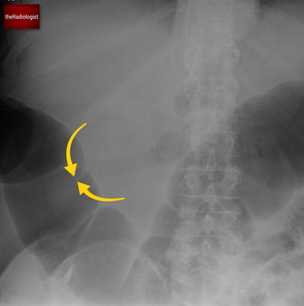
In this case instead of valvulae conniventes traversing the whole wall of the bowel which we with small bowel, we can see haustrae which we can just see at the periphery of the bowel lumen. This along with the degree of distension points us towards mechanical large bowel obstruction.
We can see haustrae which do not traverse the whole bowel wall rather than valvulae conniventes within small bowel which do – this points to large bowel dilatation.
When we look at the CT we will see diffusely dilated large bowel loops. The key here is to see whether we can find a transition point, ie is there a point where the dilated large bowel becomes collapsed.
If we follow the sigmoid colon down we will find some mural thickening and a transition point suggesting mechanical large bowel obstruction. The differential at this point lies between a malignant stricture and a benign diverticular stricture.
If we follow the colon round from the caecum we will find a transition from dilated colon to collapsed sigmoid colon. At this point there is some mural thickening. Now what are the causes of large bowel obstruction? The list is long but the most common and the most serious cause is colonic carcinoma (compare with this with small bowel obstruction where the majority of the time the aetiology is non-malignant). Other more common causes of large bowel obstruction include diverticular disease and sigmoid volvulus.
Have a look at the table below where we go through a more thorough list of the causes of large bowel obstruction in a systematic way. Breaking it down in this way in a viva can help the examiner know you are thinking systematically even if you forget a few of the causes.
| Category | Cause | Notes |
|---|---|---|
| Malignancy | Colorectal cancer | Most common cause. |
| Extrinsic compression from pelvic malignancies | Such as ovarian, prostate and bladder malignancies with associated nodal disease. | |
| Volvulus | Sigmoid volvulus | More common in elderly. |
| Caecal volvulus | Less likely to present with large bowel obstruction, can cause small bowel obstruction if patent ileocaecal valve. | |
| Inflammatory/infective | Diverticulitis | When associated with stricture formation. |
| Colitis | Inflammatory bowel disease (ie Crohn’s with stricture formation), ischaemic colitis, radiation colitis | |
| Post surgical | Anastomotic stricture | |
| Post op adhesional stricture | More commonly causes small bowel obstruction. | |
| Hernia | Inguinal, femoral, abdominal wall, incisional | Incarcerated large bowel loops with upstream dilatation. Can also cause small bowel obstruction. |
| Functional | Faecal impaction | Usually elderly, bed bound patients |
| Intussusception | Intussusception | In adults look for a lead point such as a tumour or polyp |
| Extrinsic compression | Abscess | |
| Pelvic/retroperitoneal mass |
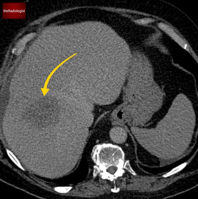
Going back to our case, with mural thickening within the sigmoid colon we have to flag that this is a possible colon cancer. What makes us more certain is the presence of low density ill defined liver lesions which represent liver metastases. Note how these do not show fluid density and are not well defined as you would expect with simple liver cysts.
The patient went on to have a colonic stent before resection of the obstructing lesion for symptom control and then chemotherapy.
There is a large ill defined low density lesion within the liver suggestive of a liver metastasis.
Colon cancer is the most common cause of large bowel obstruction with other common causes including diverticular disease and sigmoid volvulus.
Conversely most cases of small bowel obstruction are non-maignant in aetiology.
Downloaded by 10,000+ healthcare professionals.
Get 20 annotated chest X-ray signs with clear teaching points and explanations. Written by a Consultant Radiologist, this free guide is designed to help you recognise important findings more confidently when reviewing chest X-Rays.
You’ve got the framework now let’s put it to work. Dive into a related case to see these findings on real images, or explore another guide to build out your systematic approach. That’s where it really starts to stick.
A systematic approach to bowel gas pattern, obstruction and key checks
Dilated bowel loops and key review areas on CT and X-Ray
X-ray signs, differentiating from caecal volvulus and CT features
We use cookies to make this website work and to understand how it is used. You can accept all cookies, use essential cookies only or manage your preferences.