A woman in her 50s presents with persistent headache and unsteady gait. Brain MRI shows a lesion at the foramen of Monro causing obstructive hydrocephalus, in keeping with a colloid cyst.
This case shows how to read the MRI, recognise acute hydrocephalus, identify transependymal oedema and consider the key differentials.
Useful for radiology, neurology, emergency medicine and anyone improving brain MRI interpretation.
A woman in her 50s, with no significant medical history, visits her GP complaining of a persistent headache and an altered gait. Given her symptoms, she undergoes an urgent MRI of the brain. Shortly after the scan, she is admitted to the hospital.
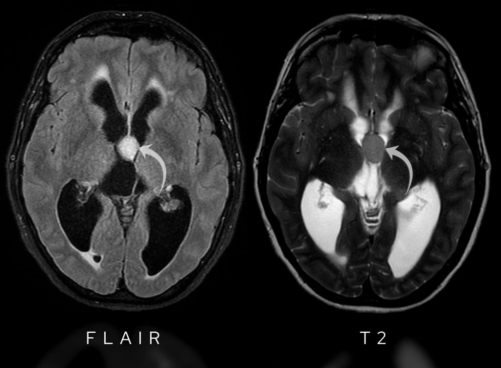
Have a look at the axial FLAIR images of the scan below. What does it show?
Tap below to open the interactive case. Then give it a few seconds to fully load.
Here is a video explanation of this case: click full screen in the bottom right corner to make it big. If you prefer though I go through this in the text explanation below.
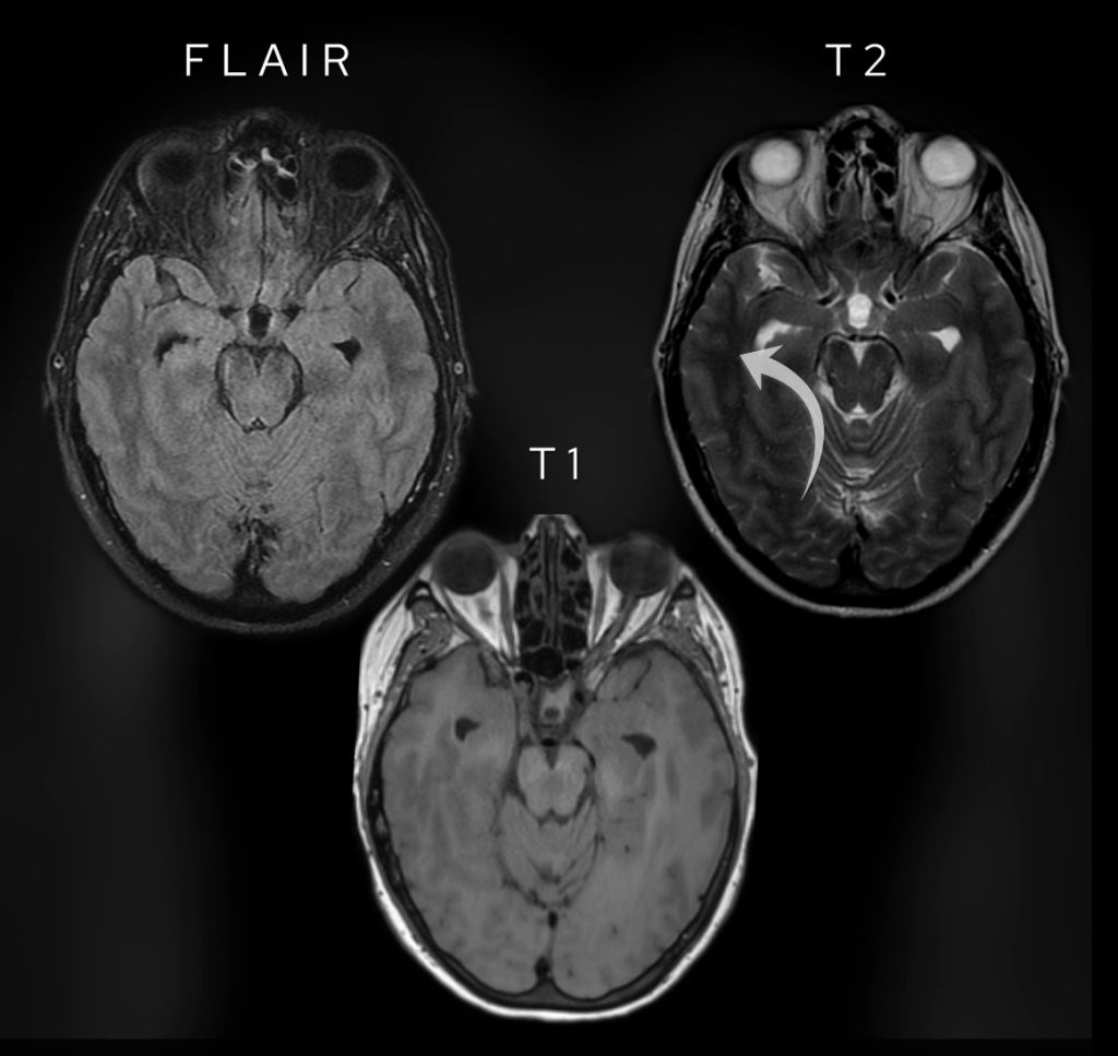
One of the key sequences used in brain imaging is FLAIR (Fluid Attenuated Inversion Recovery). It’s a variation of T2-weighted imaging but with fluid (like CSF) suppressed, making it excellent for detecting inflammation and oedema. Have a look at the images below and compare the fluid within the CSF, the grey matter and the white matter.
Note the difference between FLAIR images and T1 weighted and T2 weighted images. Fluid is dark like on T1 weighted images. Note the white matter (arrowed on the T2 image) is brighter than grey matter only on T1 weighted images.
Like on T2-weighted images, oedema shows as bright but unlike T2-weighted images fluid is dark (as can be seen on T1-weighted images). Have a look at the table below which highlights some of the key differences.
| Sequence | Fluid | White versus grey matter | Oedema | Fat |
|---|---|---|---|---|
| T1-weighted | Dark | White brighter than grey | Not seen | Bright |
| T2-weighted | Bright | White darker than grey | Bright | Bright |
| FLAIR | Dark | White darker than grey | Bright | Dark |
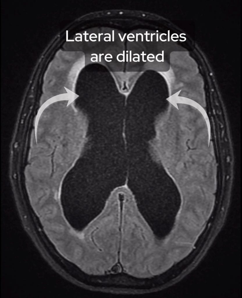
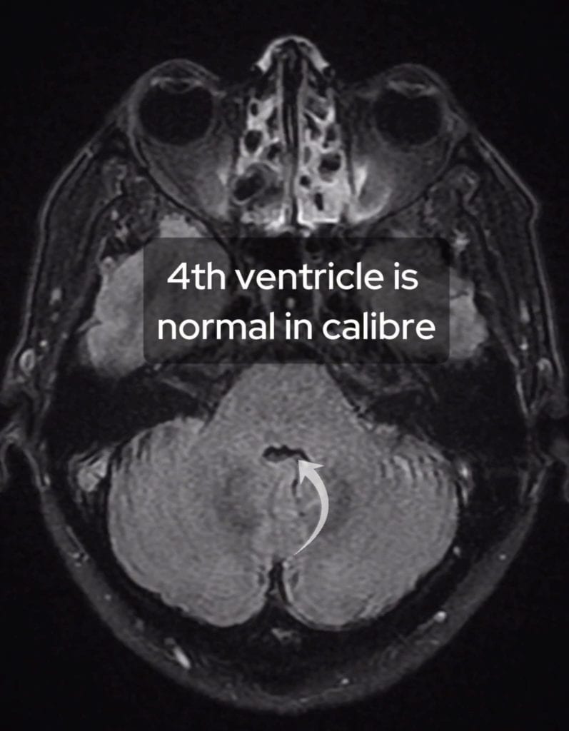
In this case, we immediately notice marked dilation of the lateral ventricles, an important clue. However, when we look further down at the fourth ventricle, it appears normal in size.
This suggests obstructive hydrocephalus, meaning something is blocking the normal flow of cerebrospinal fluid (CSF). What’s causing the obstruction?
The lateral ventricles are dilated suggesting there is hydrocephalus.
Although the lateral ventricles are dilated, the 4th ventricle is normal in calibre suggesting there is obstructive hydrocephalus.
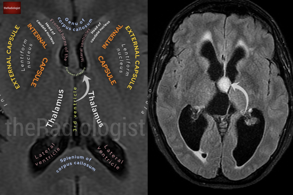
Have a look at the normal annotated brain scan below and compare it to our case.
A normal brain scan shows that the foramen of Monro connects the lateral ventricles to the third ventricle. However, in our patient’s scan, there is a well-defined lesion in the roof and anterior aspect of the third ventricle, right at the foramen of Monro.
Note the normal anatomy of the foramen of Monro on the left and the presence of an obstructing lesion on the image on the right.
Let’s examine its MRI characteristics:
FLAIR images show hyperintensity within the lesion whilst T2-weighted images show the lesion is hypointense to CSF.
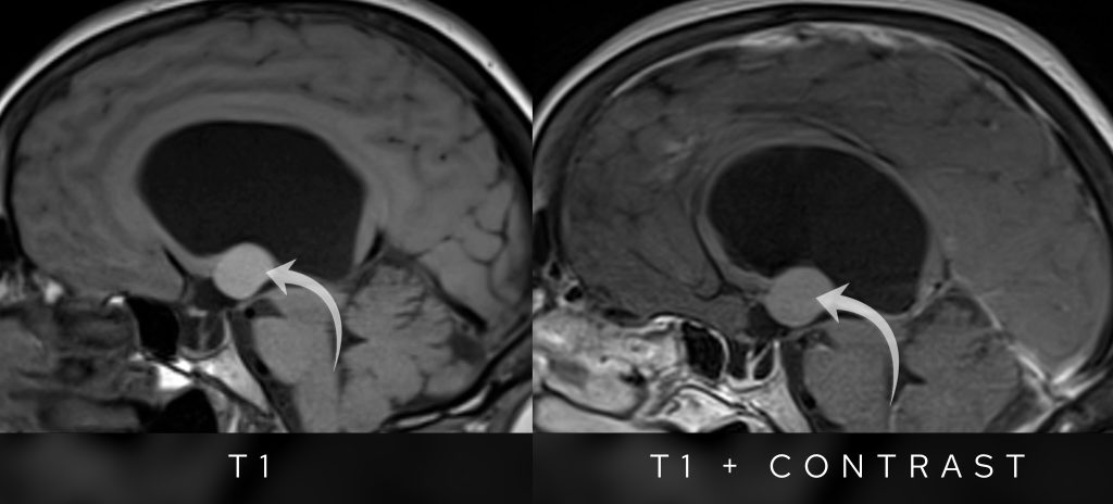
T1 pre and post contrast sagittal images show no appreciable enhancement in the lesion post intravenous gadolinium contrast.
So what is the most likely diagnosis?
The lesion is most compatible with a colloid cyst.
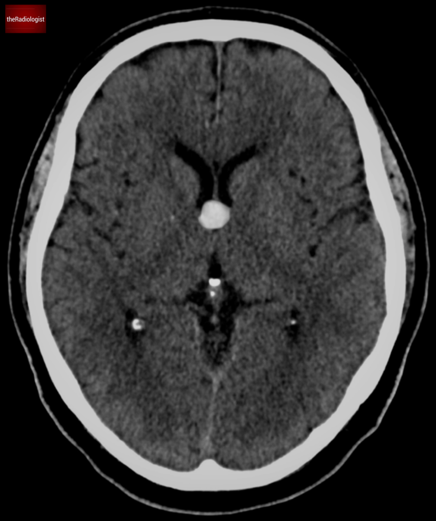
A colloid cyst is a benign epithelial-lined cyst that typically forms at the foramen of Monro. These cysts can be found incidentally on CT scans as small high-density regions in this area.
Colloid cysts usually show on CT as a high density lesion in the region of the foramen of Monro.
Why does it matter? Because of its location, even a relatively small colloid cyst can block CSF flow, leading to significant hydrocephalus and symptoms like headache, balance problems, and even sudden neurological decline.
Let’s think about what the differential in this case is.
This all points strongly toward a colloid cyst as the leading diagnosis.
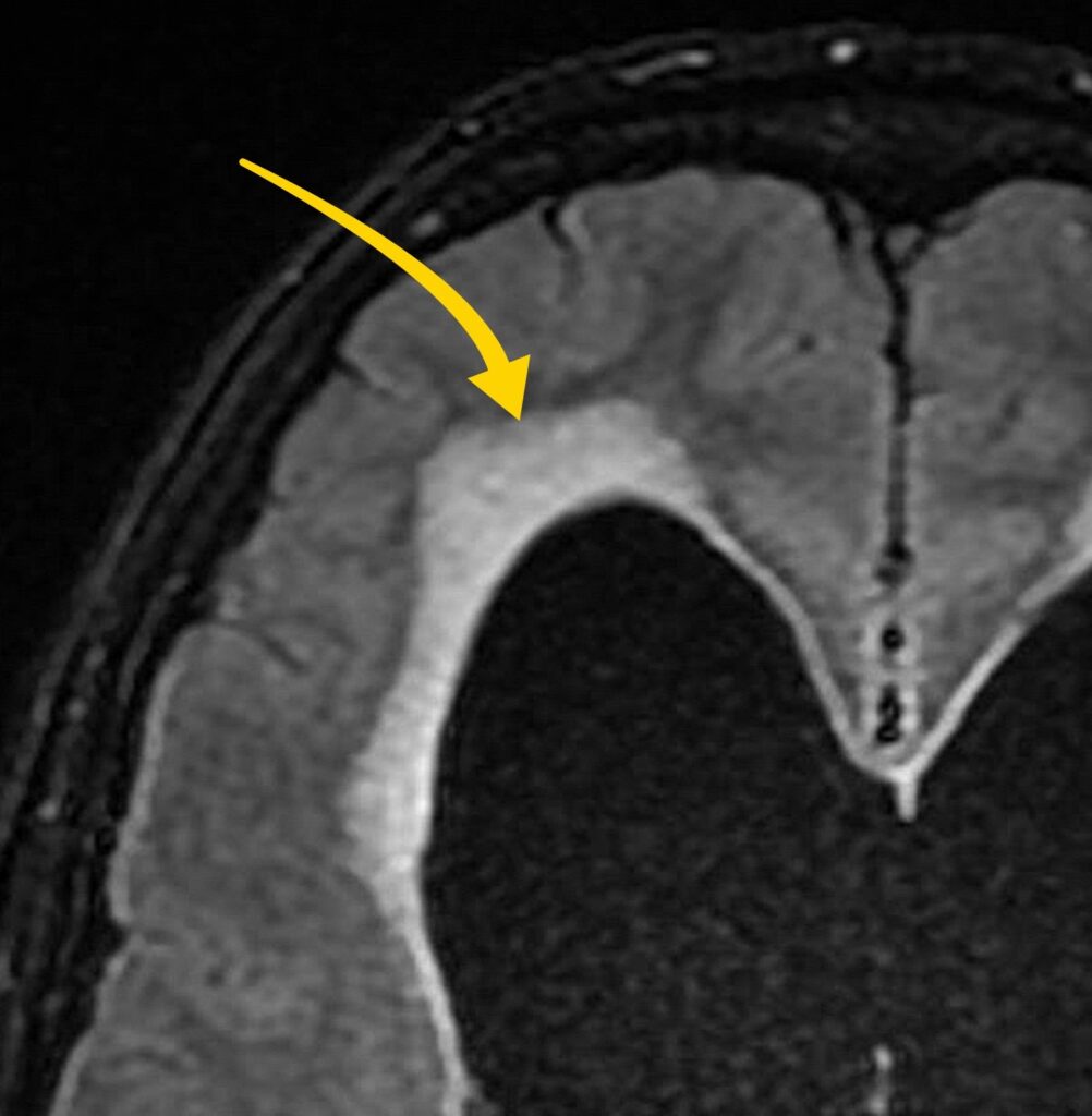
Going back to the FLAIR sequence, we see high signal surrounding the lateral ventricles—this is transependymal oedema, an important indicator of acute obstructive hydrocephalus.
What’s happening here?
This is a serious finding: it suggests acute hydrocephalus, which, if untreated, can lead to brain herniation and death.
Fluid leak in the context of acute hydrocephalus can lead to transependymal oedema and the appearance of hyperintensity on FLAIR images.
The treatment for a colloid cyst depends on:
In this case, the colloid cyst was causing life-threatening hydrocephalus, so urgent surgical resection was performed.
Once the cyst was removed, the hydrocephalus resolved, and the patient’s symptoms improved.
Here’s a summary table of the key features of colloid cysts:
| Colloid cyst facts |
|---|
| Benign cyst filled with gelatinous material |
| Typical location within the anterior third ventricle |
| Variable size |
| Commonly asymptomatic and an incidental finding |
| Can cause acute hydrocephalus/drop attack |
| On CT typically looks bright (hyperdense) |
| MRI can have variable signal characteristics |
Don’t forget to overlook the foramen of Monro for a colloid cyst which can be missed.
Transependymal oedema showing as high T2/FLAIR signal on MRI and low density on CT surrounding the ventricles can be a sign of acute hydrocephalus.
Downloaded by 10,000+ healthcare professionals.
Get 20 annotated chest X-ray signs with clear teaching points and explanations. Written by a Consultant Radiologist, this free guide is designed to help you recognise important findings more confidently when reviewing chest X-Rays.
You’ve got the framework now let’s put it to work. Dive into a related case to see these findings on real images, or explore another guide to build out your systematic approach. That’s where it really starts to stick.
CT head and CT angiogram features, review areas and pitfalls
Brain CT and MRI features, mass effect and differential diagnosis
We use cookies to make this website work and to understand how it is used. You can accept all cookies, use essential cookies only or manage your preferences.