Bronchiectasis
Learn X-Ray and CT features of bronchiectasis
Introduction
Bronchiectasis is a chronic lung condition characterised by irreversible dilatation of the bronchi. It commonly presents with persistent cough, recurrent infections and excess sputum.
From a radiology perspective, the key is recognising the imaging signs. Chest X-ray may show subtle features such as ring shadows, tram-track opacities and bronchial wall thickening while CT is the main test for diagnosis.
In this article, we go through a real-life case of bronchiectasis to illustrate the chest X-ray and CT appearances, covering the definition, causes, radiographic signs and CT patterns.
Case introduction
A man in his 20s attends his GP after struggling with recurrent chest infections. A chest X-ray is performed—do you see anything abnormal? This is a tricky one.
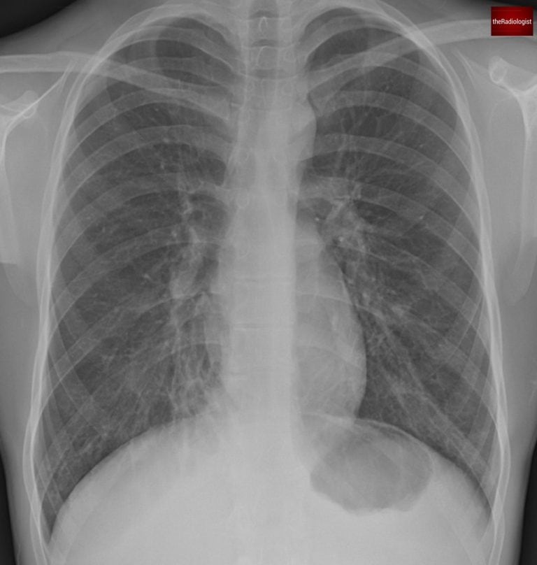
PA view of a chest X-Ray of a male in his 20s
Video explanation
Here is a video explanation of this case: click full screen in the bottom right corner to make it big. If you prefer though I go through this in the text explanation below.
Case findings
When looking at a chest X-ray for someone in their 20s, you generally expect clear lungs with no abnormalities. So even subtle abnormalities in a patient of this age should not be taken lightly.
Let’s start by having a look at the right lung near the right heart border and compare with the same area within the left lower zone.
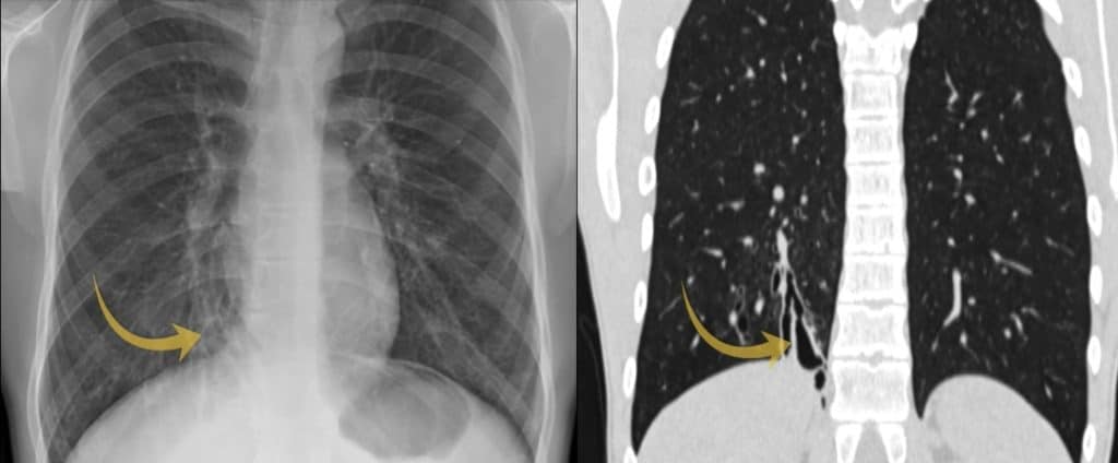
We can see the area of lung by the right heart border looks abnormal and if you look closely you can just make out a ‘ring shadow’: this literally has the appearance of a ring. Ring shadows on a chest X-ray raise the possibility of bronchiectasis. Another term used on X-Ray is ‘tram tracks’: similar to ring shadows but used when the outline is more tubular. You can see some of these above the arrowed ring shadow on the picture below.
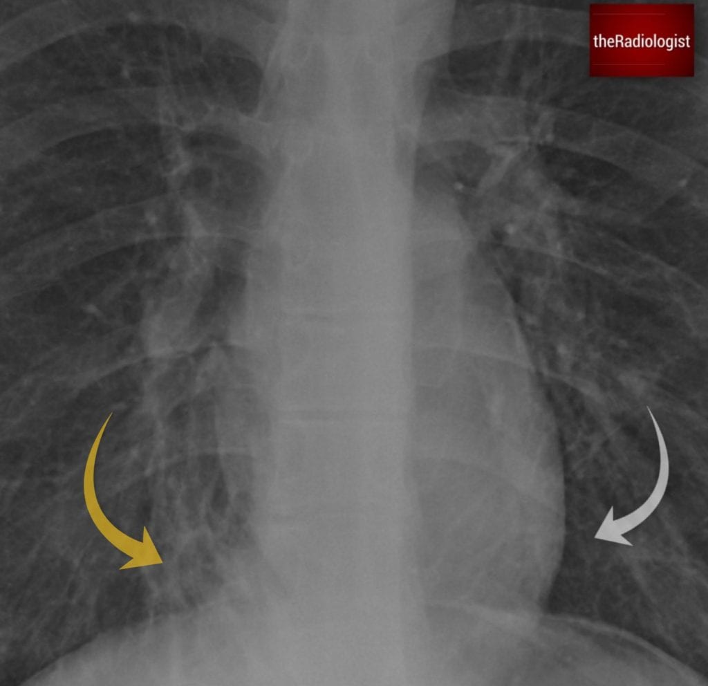
Compare the area of lung adjacent to the right (yellow arrow) and left heart border (white arrow) and you will see the region adjacent to the right heart border appears abnormal. Look closely and you will see a ‘ring shadow’ raising the possibility of bronchiectasis. Above the ring shadow are some ‘tram tracks’.
Bronchiectasis definition
Bronchiectasis is a condition where the airways become abnormally dilated and damaged. Note the airway changes in bronchiectasis are irreversible.
How do you diagnose bronchiectasis?
A CT scan is the gold standard because it lets us see the airway size and structure more clearly. Normally, the airway should be the same size or smaller than the pulmonary artery running alongside it. In bronchiectasis, the airway is larger than the adjacent pulmonary artery. On CT scans this can give something called a ‘signet ring’ sign: you can see a dot (pulmonary artery) with the ring (dilated bronchus) next to it giving the impression of a signet ring.
CT is far more sensitive than X-Ray and can show bronchial dilatation when compared with the adjacent pulmonary artery, lack of tapering and the signet ring sign as talked about above. If X-Ray doesn’t show bronchiectasis but you are suspecting it, a non contrast HRCT (high resolution CT) scan of the chest should be able to give you a diagnosis of bronchiectasis if it is present.
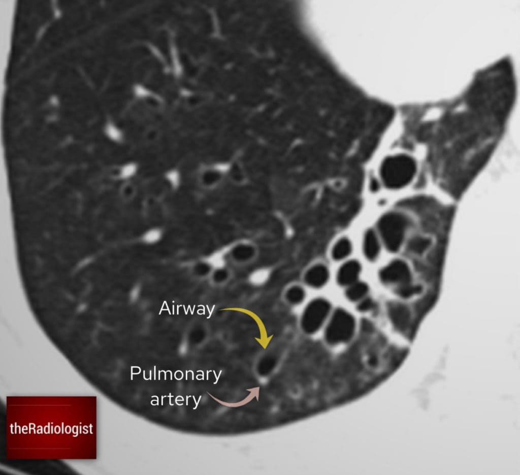
In normal circumstances the airway should be smaller or the same size as the pulmonary artery running beside it. I often think can I fit the pulmonary artery into the airway as if it were a hole – if I can’t then there is no bronchiectasis.
Causes of bronchiectasis
Bronchiectasis can sometimes happen without an obvious cause (idiopathic), but there are several things that can make someone more likely to develop it:
| Cause | Notes |
|---|---|
| Recurrent infection or serious childhood infection | Severe or repeated chest infections, especially in childhood, can damage the airways permanently. Illnesses like whooping cough or measles can lead to long-term lung damage. |
| Cystic fibrosis | This condition leads to thick mucus buildup, causing chronic infections and, ultimately, bronchiectasis. If you see bronchiectasis on a CT scan look for fatty replacement of the pancreas and this can lead you to a diagnosis of cystic fibrosis. |
| Allergic bronchopulmonary aspergillosis (ABPA) | A hypersensitivity reaction to the fungus Aspergillus, almost exclusively seen in asthmatics, causes central bronchiectasis and mucus-filled bronchi that can look like a finger-in-glove sign on CT. High-density mucus on CT? That’s a big clue that ABPA is the underlying cause. |
| Airway obstruction | If something is blocking an airway (like a tumour or foreign body), mucus builds up behind it, leading to localised bronchiectasis so look proximal to the bronchiectasis if focal on CT to see if anything is blocking the airway. |
| Other | Other causes include primary ciliary dyskinesia, immunodeficiency disorders and rheumatoid arthritis. |
Case workup
On the CT scan if we assess the right lower lobe we will see dilatation of the airways and bronchiectasis.
We can also see signs of airway inflammation:
- Bronchial wall thickening – A sign of ongoing airway inflammation.
- Groundglass centrilobular nodules – These suggest inflammation affecting the small airways, possibly infection in the context of bronchiectasis.
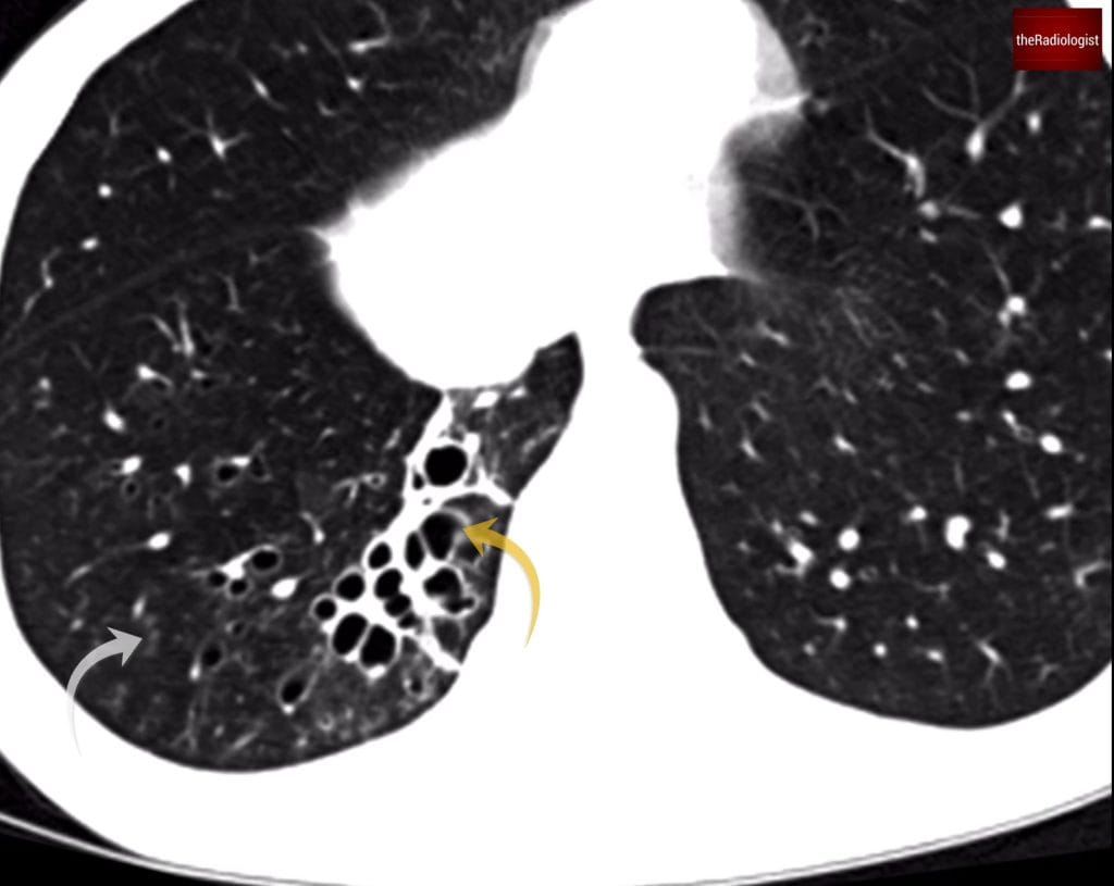
Within the posterior segment of the right lower lobe there is clear bronchiectasis (yellow arrow) and signs of airway inflammation. The grey arrow points at centrilobular ground nodules, a sign of small airway inflammation.
Types of bronchiectasis
We can describe bronchiectasis in different ways when we see it on CT. We can think of it as a spectrum.
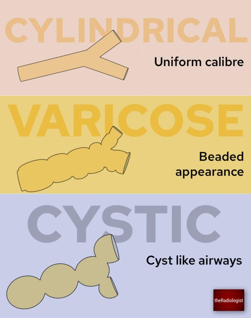
We can think of bronchiectasis as a spectrum of cylindrical, varicose and cystic bronchiectasis.
Cylindrical bronchiectasis
Airway walls look smooth and tubular. Ie they look like cylinders.
Varicose bronchiectasis
The airways look beaded, like a string of pearls.
Cystic bronchiectasis
The most severe form, where airways look like clusters of cysts, sometimes described as a ‘bunch of grapes’ and this is what we can see in our case. Cystic bronchiectasis can mimic cystic lung disease or honeycombing as seen in lung fibrosis.
How can we tell the difference? with cystic bronchiectasis the cysts will follow the course of the airways and you should be able to trace the cysts back to the proximal airway.
Cystic bronchiectasis is thought to resemble a ‘bunch of grapes’.
Reflection
Now that we’ve analysed the CT, let’s go back to the original chest X-ray. The ring shadows represented dilated airways seen end-on and are something to look out for on X-Ray.
The area of abnormality on the chest X-Ray correlates with the area of cystic bronchiectasis seen on CT.
KEY POINTS
X-Rays in young patients in particular should be ‘clean’ – if you see an abnormality you need to be able to explain it.
Ring shadows may represent underlying bronchiectasis for which CT is the gold standard for diagnosis.
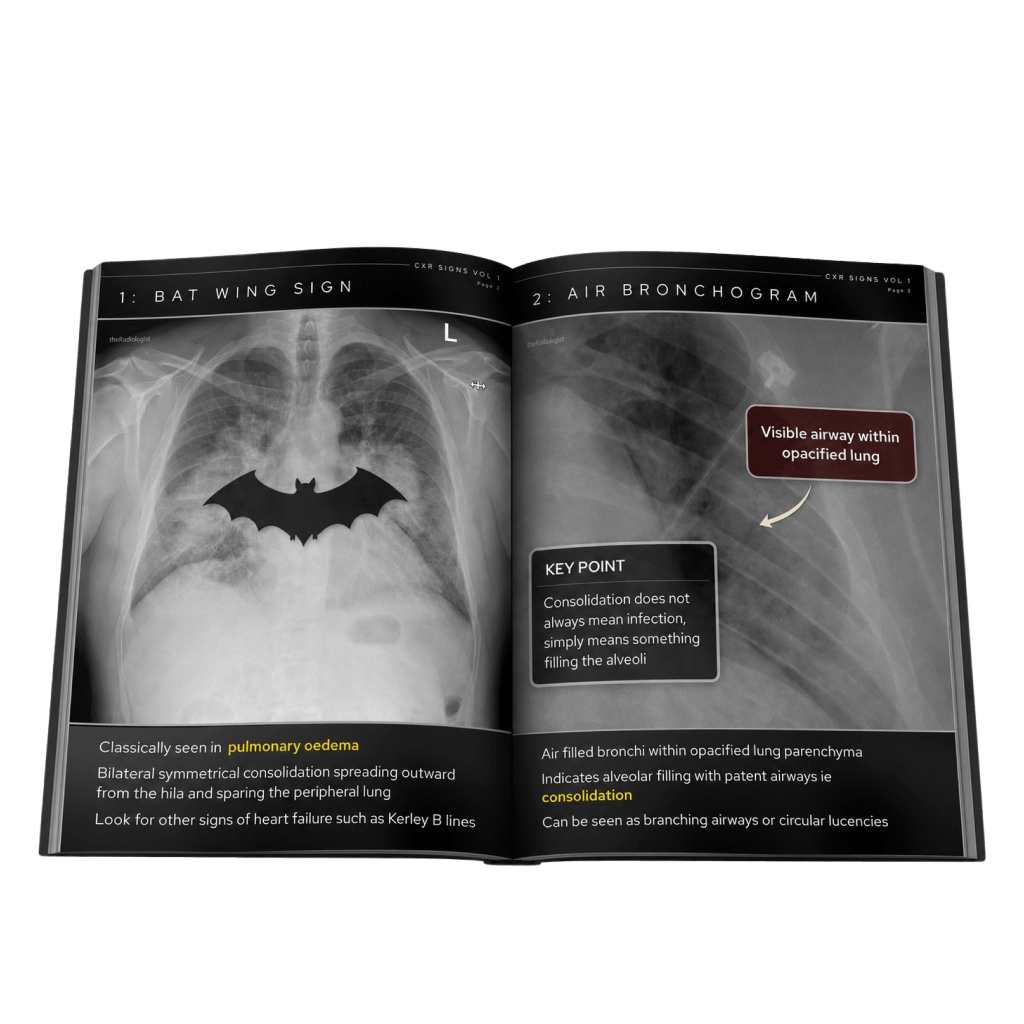
Free guide: 20 Chest X-Ray signs you need to recognise
Downloaded by 10,000+ healthcare professionals.
Get 20 annotated chest X-ray signs with clear teaching points and explanations. Written by a Consultant Radiologist, this free guide is designed to help you recognise important findings more confidently when reviewing chest X-Rays.