CT head features, basal ganglia anatomy and causes
When it comes to CT head interpretation, recognising an intracerebral haemorrhage quickly and accurately is vital. In this case, we focus on a deep bleed in the basal ganglia.
You’ll learn how to spot key CT features like mass effect and intraventricular blood, and understand the typical causes of basal ganglia haemorrhage.
A male in his 50s presents to the Emergency Department with loss of consciousness. What does the CT scan show?
Tap below to open the interactive case. Then give it a few seconds to fully load.
Here is a video explanation of this case: click full screen in the bottom right corner to make it big. If you prefer though I go through this in the text explanation below.
Let’s take a closer look at this CT head scan: there’s something clearly abnormal on the left side of the brain. We perform most acute CT head scans without IV contrast as haemorrhage appears bright and we don’t want bright IV contrast to mask this.
What Normally Appears Bright on a CT Head?
Some structures naturally show up as bright (hyperdense) on CT scans:
However if you can’t explain high density by calcification or a normal structure, you need to seriously consider haemorrhage.
Once you’ve found haemorrhage you’ll want to try and locate where it is. Haemorrhage on a brain CT can be typically be in one of the following compartments:
The management of each can be different and the causes can also vary. Remember that you can get blood within more than one compartment especially in cases of trauma.
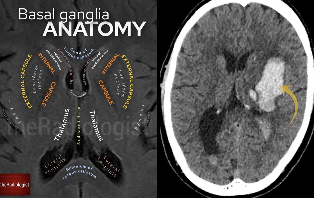
Now, looking at our patient’s scan, the bleed is clearly centered in the lentiform nucleus and more specifically, the putamen, which together with the globus pallidus makes up the lentiform nucleus. This makes it an intraparenchymal bleed.
The bleed is centred on the left lentiform nucleus and more specifically the putamen.
While a tumour or underlying lesion could be a possibility, the most common cause in this location is high blood pressure (hypertension).
Chronic hypertension can cause tiny, weak-walled microaneurysms called Charcot–Bouchard aneurysms, which can rupture and cause bleeding. These tend to happen in specific locations, and the basal ganglia—especially the putamen—is one of the most common sites. Other typical locations include:
A deep hemorrhage in the brain is never good news, but there are certain signs that make this case even more worrisome:
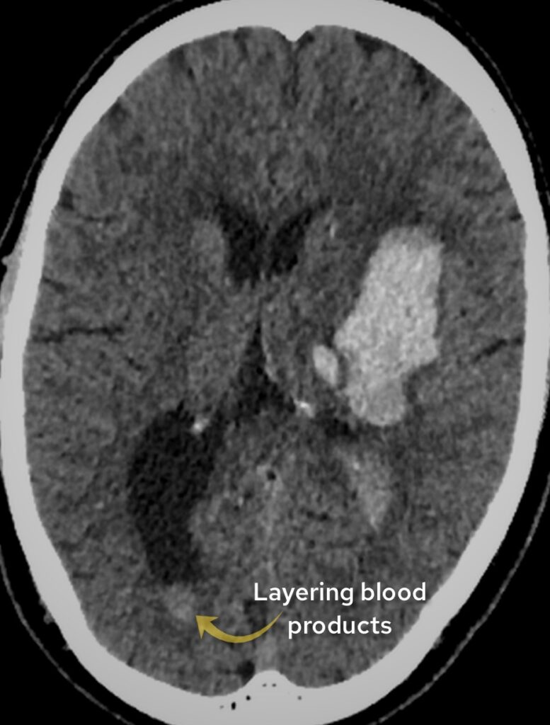
Having intraventricular hemorrhage (blood in the ventricles) is linked to a worse prognosis and worsened patient outcomes.
There is mass effect: there is effacement of the left lateral ventricle caused by the haemorrhage.
Blood is seen within a dependent region of the right lateral ventricle: always a key review area.
Deep brain haemorrhage within the basal ganglia, thalamus, pons and cerebellum are highly suggestive of a hypertensive bleed.
Once you see this look for secondary signs such as mass effect and intraventricular haemorrhage.
Downloaded by 10,000+ healthcare professionals.
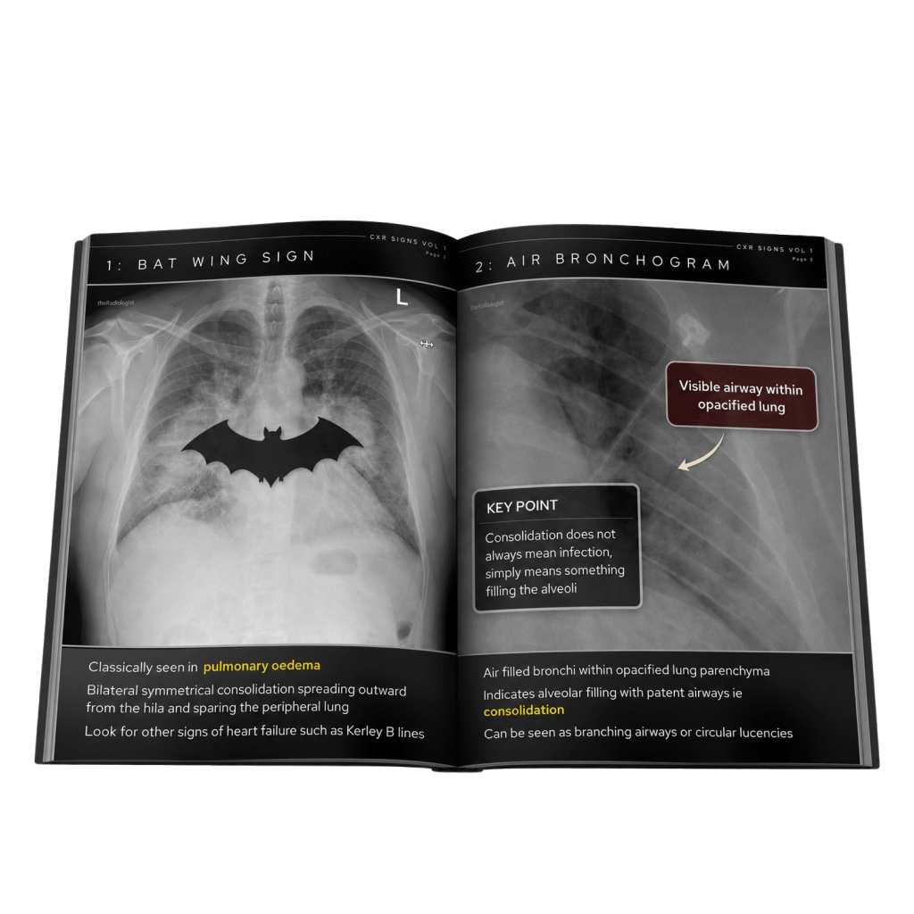
Get 20 annotated chest X-ray signs with clear teaching points and explanations. Written by a Consultant Radiologist, this free guide is designed to help you recognise important findings more confidently when reviewing chest X-Rays.
You’ve got the framework now let’s put it to work. Dive into a related case to see these findings on real images, or explore another guide to build out your systematic approach. That’s where it really starts to stick.
CT head and CT angiogram features, review areas and pitfalls
Review the anatomy, early signs of an MCA infarct and CT angiogram findings
Brain CT and MRI features, mass effect and differential diagnosis
We use cookies to make this website work and to understand how it is used. You can accept all cookies, use essential cookies only or manage your preferences.