Fat pads might seem like a small detail, but they can make a big difference in diagnosing elbow injuries. In this case, we look at how to assess the anterior and posterior fat pads on an elbow X-ray, even when the image isn’t perfect.
You’ll learn the key fat pad rules, why their position matters, and how they can point to an occult fracture like a subtle radial head break. A quick, high-yield guide for spotting hidden trauma on elbow films.
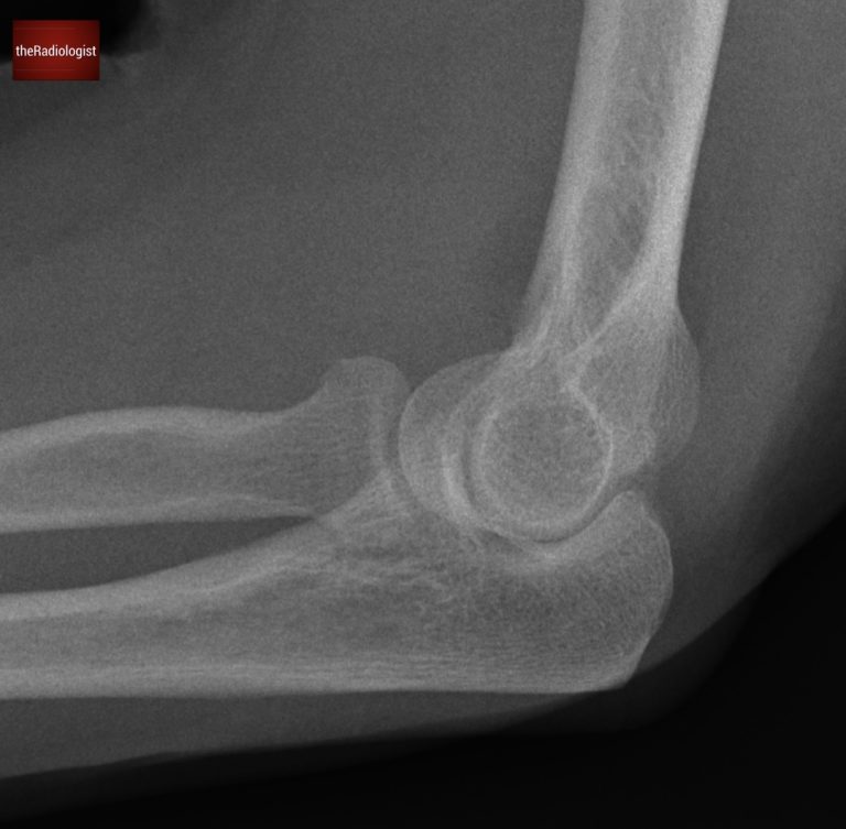
Have a look at this case of elbow trauma. The projection is suboptimal but regardless, there’s a clear abnormality. The key to interpreting this film lies in recognizing the significance of fat pads. Let’s break it down.
Lateral Elbow X-Ray
Video explanation
Here is a video explanation of this case: click full screen in the bottom right corner to make it big. If you prefer though I go through this in the text explanation below.
Normal elbow fat pad anatomy
To understand what’s abnormal, let’s first review normal fat pad anatomy:
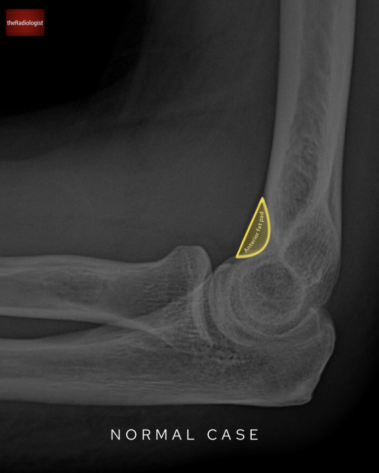
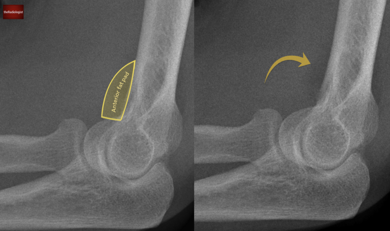
1. Anterior fat pad
Normally lives within the coronoid fossa.
May not be visible at all, or if present, appears as a thin stripe parallel to the anterior humerus.
Anterior fat pad on a normal elbow X-Ray.
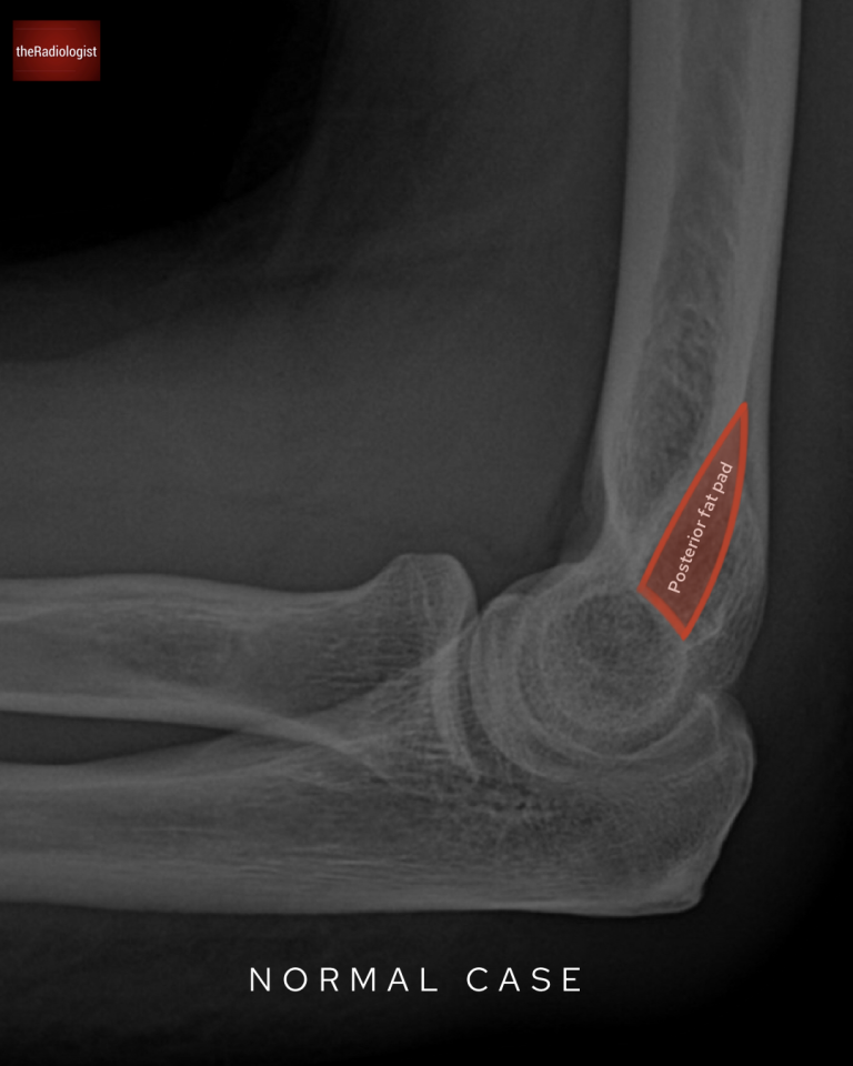
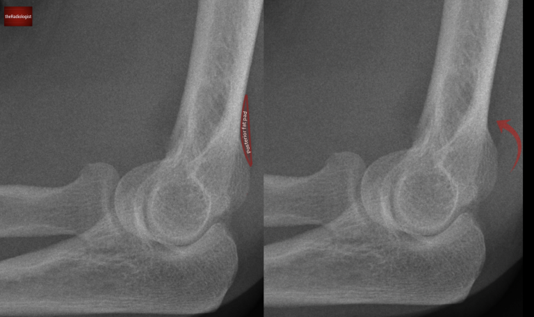
2. Posterior fat pad
Resides within the olecranon fossa.
Normally hidden by bone and not visible on X-ray.
Posterior fat pad on a normal elbow X-Ray: this is normally obscured.
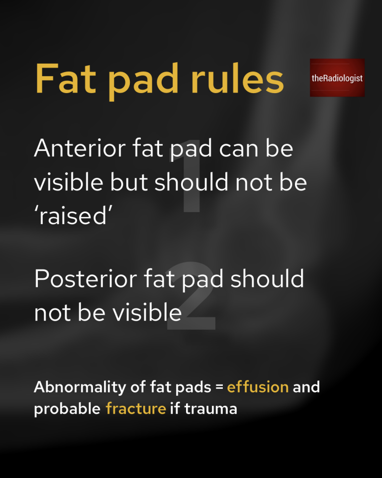
Fat pad rules
There are two essential rules to remember when assessing fat pads on an elbow X-ray:
The anterior fat pad can be visible but should not be raised.
The posterior fat pad should not be visible at all.
The fat pad rules.
Case findings
Going back to our case, the alignment isn’t the best on our X-Ray in that the capitellum and trochlea do not overlap each other. However sometimes we have to deal with suboptimal images and still apply the rules we know. Let’s apply our fat pad rules:
Instead of a thin stripe the anterior fat pad is clearlyraised, signaling something abnormal. Now let’s have a look for a posterior fat pad.
A ‘raised’ abnormal anterior fat pad can be seen.
The posterior fat pad is visible in this case when in normal circumstances it should be obscured.
What does this mean? The presence of abnormal fat pads indicates an elbow joint effusion. In the context of trauma, this strongly suggests an intra-articular fracture.
We can see a posterior fat pad which in normal circumstances should be obscured like the case above.
Assessing for fracture
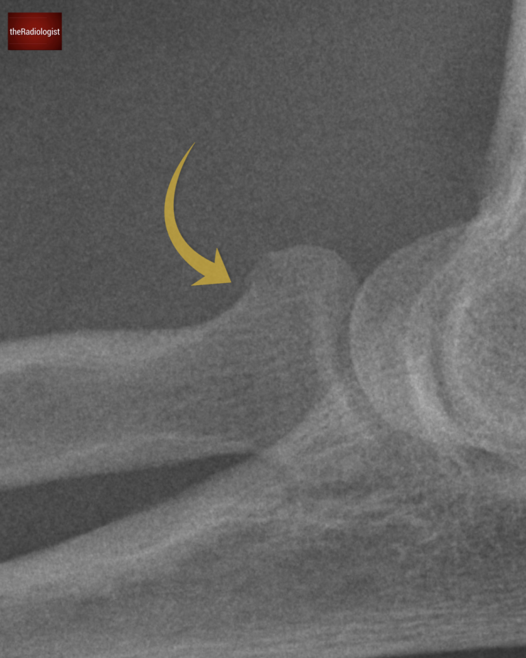
In adults, the most common elbow fracture is a radial head fracture, so that’s the first place to check.
On closer inspection, you can spot a subtle cortical break in the radial head.
Although it’s subtle, the presence of abnormal fat pads confirm that this is indeed a fracture.
Looking closely we can see a subtle fracture at the radial head.
KEY POINT
Don’t forget the fat pad rules!
The anterior fat pad can be visible but not raised.